


 癌症的基本特征包括细胞增殖、血管生成、迁移、凋亡逃避机制和细胞永生等。找到癌症发生过程中这些通路的关键标记物和对应的抗体用于检测至关重要。
 为您推荐一个泛素化位点预测神器——泛素化分析工具,可以为您的蛋白的泛素化位点作出预测和评分。
 细胞自噬受体图形绘图工具为你的蛋白的细胞受体结合位点作出预测和评分,识别结合到自噬通路中的蛋白是非常重要的,便于让我们理解自噬在正常生理、病理过程中的作用,如发育、细胞分化、神经退化性疾病、压力条件下、感染和癌症。
ITK Antibody
Purified Mouse Monoclonal Antibody
- 产品详情
- 实验流程
Application
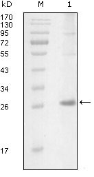
| WB, E |
|---|---|
| Primary Accession | Q08881 |
| Reactivity | Human |
| Host | Mouse |
| Clonality | Monoclonal |
| Clone Names | 5G12C4 |
| Isotype | IgG1 |
| Calculated MW | 71831 Da |
| Description | ITK: IL2-inducible T-cell kinase. This gene encodes an intracellular tyrosine kinase expressed in T-cells. The protein contains both SH2 and SH3 domains which are often found in intracellular kinases. It is thought to play a role in T-cell proliferation and differentiation. |
| Immunogen | Purified recombinant fragment of ITK (aa2-110) expressed in E. Coli. |
| Formulation | Ascitic fluid containing 0.03% sodium azide. |
| Gene ID | 3702 |
|---|---|
| Other Names | Tyrosine-protein kinase ITK/TSK, 2.7.10.2, Interleukin-2-inducible T-cell kinase, IL-2-inducible T-cell kinase, Kinase EMT, T-cell-specific kinase, Tyrosine-protein kinase Lyk, ITK, EMT, LYK |
| Dilution | WB~~1/500 - 1/2000 E~~N/A |
| Storage | Maintain refrigerated at 2-8°C for up to 6 months. For long term storage store at -20°C in small aliquots to prevent freeze-thaw cycles. |
| Precautions | ITK Antibody is for research use only and not for use in diagnostic or therapeutic procedures. |
| Name | ITK |
|---|---|
| Synonyms | EMT, LYK |
| Function | Tyrosine kinase that plays an essential role in regulation of the adaptive immune response. Regulates the development, function and differentiation of conventional T-cells and nonconventional NKT-cells. When antigen presenting cells (APC) activate T-cell receptor (TCR), a series of phosphorylation lead to the recruitment of ITK to the cell membrane, in the vicinity of the stimulated TCR receptor, where it is phosphorylated by LCK. Phosphorylation leads to ITK autophosphorylation and full activation. Once activated, phosphorylates PLCG1, leading to the activation of this lipase and subsequent cleavage of its substrates. In turn, the endoplasmic reticulum releases calcium in the cytoplasm and the nuclear activator of activated T-cells (NFAT) translocates into the nucleus to perform its transcriptional duty. Phosphorylates 2 essential adapter proteins: the linker for activation of T-cells/LAT protein and LCP2. Then, a large number of signaling molecules such as VAV1 are recruited and ultimately lead to lymphokine production, T-cell proliferation and differentiation (PubMed:12186560, PubMed:12682224, PubMed:21725281). Required for TCR-mediated calcium response in gamma-delta T-cells, may also be involved in the modulation of the transcriptomic signature in the Vgamma2-positive subset of immature gamma-delta T-cells (By similarity). Phosphorylates TBX21 at 'Tyr-530' and mediates its interaction with GATA3 (By similarity). |
| Cellular Location | Cytoplasm. Nucleus {ECO:0000250|UniProtKB:Q03526}. Note=Localizes in the vicinity of cell surface receptors in the plasma membrane after receptor stimulation |
| Tissue Location | T-cell lines and natural killer cell lines. |
Research Areas
For Research Use Only. Not For Use In Diagnostic Procedures.
Application Protocols
Provided below are standard protocols that you may find useful for product applications.
REFERENCES
1. Int Immunol. 2001 Oct;13(10):1265-74. 2. Biochemistry. 2002 Aug 27;41(34):10732-40. 3. J Allergy Clin Immunol. 2005 Sep;116(3):650-6.
终于等到您。ABCEPTA(百远生物)抗体产品。
点击下方“我要评价 ”按钮提交您的反馈信息,您的反馈和评价是我们最宝贵的财富之一,
我们将在1-3个工作日内处理您的反馈信息。
如有疑问,联系:0512-88856768 tech-china@abcepta.com.
¥ 1,500.00
Cat# AO1179a


