


 癌症的基本特征包括细胞增殖、血管生成、迁移、凋亡逃避机制和细胞永生等。找到癌症发生过程中这些通路的关键标记物和对应的抗体用于检测至关重要。
 为您推荐一个泛素化位点预测神器——泛素化分析工具,可以为您的蛋白的泛素化位点作出预测和评分。
 细胞自噬受体图形绘图工具为你的蛋白的细胞受体结合位点作出预测和评分,识别结合到自噬通路中的蛋白是非常重要的,便于让我们理解自噬在正常生理、病理过程中的作用,如发育、细胞分化、神经退化性疾病、压力条件下、感染和癌症。
FGR Antibody
Purified Mouse Monoclonal Antibody
- 产品详情
- 实验流程
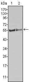
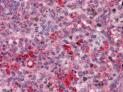
Application
| WB, IHC, E |
|---|---|
| Primary Accession | P09769 |
| Reactivity | Human, Mouse |
| Host | Mouse |
| Clonality | Monoclonal |
| Clone Names | 6G2 |
| Isotype | IgG1 |
| Calculated MW | 59479 Da |
| Description | FGR: Gardner-Rasheed feline sarcoma viral (v-fgr) oncogene homolog, also known as SRC2; c-fgr; c-src2; FLJ43153; MGC75096; p55c-fgr; p58c-fgr. It is a member of the Src family of protein tyrosine kinases (PTKs). The encoded protein contains N-terminal sites for myristylation and palmitylation, a PTK domain, and SH2 and SH3 domains which are involved in mediating protein-protein interactions with phosphotyrosine-containing and proline-rich motifs, respectively. The protein localizes to plasma membrane ruffles, and functions as a negative regulator of cell migration and adhesion triggered by the beta-2 integrin signal transduction pathway. Infection with Epstein-Barr virus results in the overexpression of this gene. Multiple alternatively spliced variants, encoding the same protein, have been identified. |
| Immunogen | Purified recombinant fragment of human FGR expressed in E. Coli. |
| Formulation | Ascitic fluid containing 0.03% sodium azide. |
| Gene ID | 2268 |
|---|---|
| Other Names | Tyrosine-protein kinase Fgr, 2.7.10.2, Gardner-Rasheed feline sarcoma viral (v-fgr) oncogene homolog, Proto-oncogene c-Fgr, p55-Fgr, p58-Fgr, p58c-Fgr, FGR, SRC2 |
| Dilution | WB~~1/500 - 1/2000 IHC~~1/200 - 1/1000 E~~N/A |
| Storage | Maintain refrigerated at 2-8°C for up to 6 months. For long term storage store at -20°C in small aliquots to prevent freeze-thaw cycles. |
| Precautions | FGR Antibody is for research use only and not for use in diagnostic or therapeutic procedures. |
| Name | FGR |
|---|---|
| Synonyms | SRC2 |
| Function | Non-receptor tyrosine-protein kinase that transmits signals from cell surface receptors devoid of kinase activity and contributes to the regulation of immune responses, including neutrophil, monocyte, macrophage and mast cell functions, cytoskeleton remodeling in response to extracellular stimuli, phagocytosis, cell adhesion and migration. Promotes mast cell degranulation, release of inflammatory cytokines and IgE-mediated anaphylaxis. Acts downstream of receptors that bind the Fc region of immunoglobulins, such as MS4A2/FCER1B, FCGR2A and/or FCGR2B. Acts downstream of ITGB1 and ITGB2, and regulates actin cytoskeleton reorganization, cell spreading and adhesion. Depending on the context, activates or inhibits cellular responses. Functions as a negative regulator of ITGB2 signaling, phagocytosis and SYK activity in monocytes. Required for normal ITGB1 and ITGB2 signaling, normal cell spreading and adhesion in neutrophils and macrophages. Functions as a positive regulator of cell migration and regulates cytoskeleton reorganization via RAC1 activation. Phosphorylates SYK (in vitro) and promotes SYK-dependent activation of AKT1 and MAP kinase signaling. Phosphorylates PLD2 in antigen-stimulated mast cells, leading to PLD2 activation and the production of the signaling molecules lysophosphatidic acid and diacylglycerol. Promotes activation of PIK3R1. Phosphorylates FASLG, and thereby regulates its ubiquitination and subsequent internalization. Phosphorylates ABL1. Promotes phosphorylation of CBL, CTTN, PIK3R1, PTK2/FAK1, PTK2B/PYK2 and VAV2. Phosphorylates HCLS1 that has already been phosphorylated by SYK, but not unphosphorylated HCLS1. Together with CLNK, it acts as a negative regulator of natural killer cell-activating receptors and inhibits interferon-gamma production (By similarity). |
| Cellular Location | Cell membrane; Lipid-anchor; Cytoplasmic side. Cell membrane; Peripheral membrane protein; Cytoplasmic side. Cell projection, ruffle membrane. Cytoplasm, cytosol. Cytoplasm, cytoskeleton. Mitochondrion inner membrane. Mitochondrion intermembrane space. Note=Detected in mitochondrial intermembrane space and at inner membranes (By similarity). Colocalizes with actin fibers at membrane ruffles. Detected at plasma membrane lipid rafts |
| Tissue Location | Detected in neutrophils, monocytes and natural killer cells (at protein level). Detected in monocytes and large lymphocytes. |
Research Areas
For Research Use Only. Not For Use In Diagnostic Procedures.
Application Protocols
Provided below are standard protocols that you may find useful for product applications.
REFERENCES
1. Immunity. 2005 Feb;22(2):235-46. 2. Biochemistry. 2007 Oct 2;46(39):11023-32.
终于等到您。ABCEPTA(百远生物)抗体产品。
点击下方“我要评价 ”按钮提交您的反馈信息,您的反馈和评价是我们最宝贵的财富之一,
我们将在1-3个工作日内处理您的反馈信息。
如有疑问,联系:0512-88856768 tech-china@abcepta.com.


