


 癌症的基本特征包括细胞增殖、血管生成、迁移、凋亡逃避机制和细胞永生等。找到癌症发生过程中这些通路的关键标记物和对应的抗体用于检测至关重要。
 为您推荐一个泛素化位点预测神器——泛素化分析工具,可以为您的蛋白的泛素化位点作出预测和评分。
 细胞自噬受体图形绘图工具为你的蛋白的细胞受体结合位点作出预测和评分,识别结合到自噬通路中的蛋白是非常重要的,便于让我们理解自噬在正常生理、病理过程中的作用,如发育、细胞分化、神经退化性疾病、压力条件下、感染和癌症。
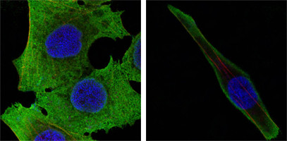
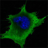
WNT5A Antibody
Purified Mouse Monoclonal Antibody
- 产品详情
- 实验流程
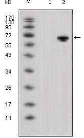
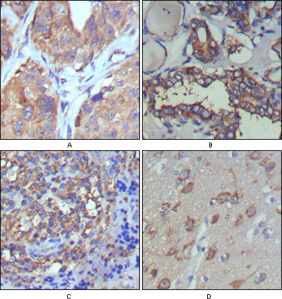
Application
| WB, IHC, E |
|---|---|
| Primary Accession | P41221 |
| Reactivity | Human |
| Host | Mouse |
| Clonality | Monoclonal |
| Clone Names | 3D10 |
| Isotype | IgG1 |
| Calculated MW | 42339 Da |
| Description | WNT5A: wingless-type MMTV integration site family, member 5A. Entrez Protein: NP_003383. The WNT gene family consists of structurally related genes which encode secreted signaling proteins. These proteins have been implicated in oncogenesis and in several developmental processes, including regulation of cell fate and patterning during embryogenesis. This gene is a member of the WNT gene family. It encodes a protein which shows 98%, 98% and 87% amino acid identity to the mouse, rat and the xenopus Wnt5A protein, respectively. The experiments performed in Xenopus laevis embryos identified that human frizzled-5 (hFz5) is the receptor for the Wnt5A ligand and the Wnt5A/hFz5 signaling mediates axis induction. |
| Immunogen | Purified recombinant fragment of WNT5A expressed in E. Coli. |
| Formulation | Ascitic fluid containing 0.03% sodium azide. |
| Gene ID | 7474 |
|---|---|
| Other Names | Protein Wnt-5a, WNT5A |
| Dilution | WB~~1/500 - 1/2000 IHC~~1/200 - 1/1000 E~~N/A |
| Storage | Maintain refrigerated at 2-8°C for up to 6 months. For long term storage store at -20°C in small aliquots to prevent freeze-thaw cycles. |
| Precautions | WNT5A Antibody is for research use only and not for use in diagnostic or therapeutic procedures. |
| Name | WNT5A |
|---|---|
| Function | Ligand for members of the frizzled family of seven transmembrane receptors. Can activate or inhibit canonical Wnt signaling, depending on receptor context. In the presence of FZD4, activates beta-catenin signaling. In the presence of ROR2, inhibits the canonical Wnt pathway by promoting beta-catenin degradation through a GSK3-independent pathway which involves down-regulation of beta- catenin-induced reporter gene expression (By similarity). Suppression of the canonical pathway allows chondrogenesis to occur and inhibits tumor formation. Stimulates cell migration. Decreases proliferation, migration, invasiveness and clonogenicity of carcinoma cells and may act as a tumor suppressor (PubMed:15735754). Mediates motility of melanoma cells (PubMed:17426020). Required during embryogenesis for extension of the primary anterior-posterior axis and for outgrowth of limbs and the genital tubercle. Inhibits type II collagen expression in chondrocytes (By similarity). |
| Cellular Location | Secreted, extracellular space, extracellular matrix. Secreted |
| Tissue Location | Expression is increased in differentiated thyroid carcinomas compared to normal thyroid tissue and anaplastic thyroid tumors where expression is low or undetectable. Expression is found in thyrocytes but not in stromal cells (at protein level) (PubMed:15735754). Detected in neonate heart and lung (PubMed:8288227) |
Research Areas
For Research Use Only. Not For Use In Diagnostic Procedures.
Application Protocols
Provided below are standard protocols that you may find useful for product applications.
REFERENCES
1. Cancer Res. 2008 Jul 15;68(14):5785-94. 2. Proc Natl Acad Sci U S A. 2009 Mar 10;106(10):3919-24.
终于等到您。ABCEPTA(百远生物)抗体产品。
点击下方“我要评价 ”按钮提交您的反馈信息,您的反馈和评价是我们最宝贵的财富之一,
我们将在1-3个工作日内处理您的反馈信息。
如有疑问,联系:0512-88856768 tech-china@abcepta.com.
¥ 1,500.00
Cat# AO1280a


