


 癌症的基本特征包括细胞增殖、血管生成、迁移、凋亡逃避机制和细胞永生等。找到癌症发生过程中这些通路的关键标记物和对应的抗体用于检测至关重要。
 为您推荐一个泛素化位点预测神器——泛素化分析工具,可以为您的蛋白的泛素化位点作出预测和评分。
 细胞自噬受体图形绘图工具为你的蛋白的细胞受体结合位点作出预测和评分,识别结合到自噬通路中的蛋白是非常重要的,便于让我们理解自噬在正常生理、病理过程中的作用,如发育、细胞分化、神经退化性疾病、压力条件下、感染和癌症。
CD80 Antibody
Purified Mouse Monoclonal Antibody
- 产品详情
- 实验流程
Application
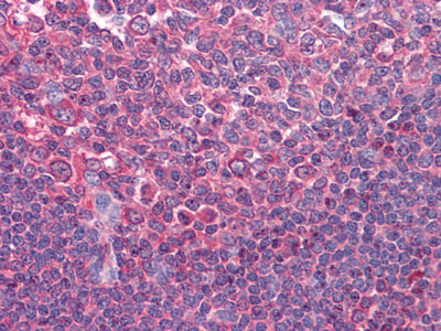
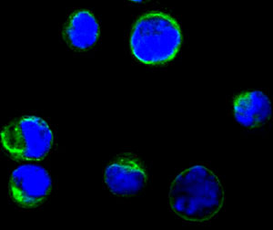
| IHC, ICC, E |
|---|---|
| Primary Accession | P33681 |
| Reactivity | Human |
| Host | Mouse |
| Clonality | Monoclonal |
| Clone Names | 2A2 |
| Isotype | IgG1 |
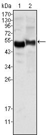
| Calculated MW | 33048 Da |
| Description | The protein CD80 (Cluster of Differentiation 80) is a molecule found on activated B cells and monocytes which provides a costimulatory signal necessary for T cell activation and survival. It is also known as B7.1. Its principal mode of action is by binding to CD28. Along with CD86, these molecules provide the necessary stimuli to prime T cells against antigens presented by antigen-presenting cells. CD80 and CD86 also bind to CTLA-4, a cell surface molecule expressed on activated T cells. Interactions between CD80 or CD86 with CTLA-4 decrease the response of T cells. Mouse research by scientists at Emory University showed that estrogen-related bone loss is linked to recently discovered pathways involving various proteins, such as CD80 and other functions. In a nutshell, reactive oxygen stimulates dendritic cells, which activate other immune cells to up-regulate production of CD80, the molecule co-responsible for T cell activation."When this pathway is activated, it leads to increased T cell TNF production and ultimately to bone loss."In turn, T cells produce a protein, Tumor Necrosis Factor, which increases the formation of osteoclasts in rodents and humans. Osteoclasts cause minerals to be released from the bone, so that calcium is taken into the bloodstream to be used for other functions of the body. Osteoclast differentiation is inhibited by osteoprotegerin; Estrogen stimulates osteoprotegerin production. |
| Immunogen | Purified recombinant fragment of CD80 expressed in E. Coli. |
| Formulation | Ascitic fluid containing 0.03% sodium azide. |
| Gene ID | 941 |
|---|---|
| Other Names | T-lymphocyte activation antigen CD80, Activation B7-1 antigen, BB1, CTLA-4 counter-receptor B7.1, B7, CD80, CD80, CD28LG, CD28LG1, LAB7 |
| Dilution | IHC~~1/200 - 1/1000 ICC~~N/A E~~N/A |
| Storage | Maintain refrigerated at 2-8°C for up to 6 months. For long term storage store at -20°C in small aliquots to prevent freeze-thaw cycles. |
| Precautions | CD80 Antibody is for research use only and not for use in diagnostic or therapeutic procedures. |
| Name | CD80 |
|---|---|
| Synonyms | CD28LG, CD28LG1, LAB7 |
| Function | Costimulatory molecule that belongs to the immunoglobulin superfamily that plays an important role in T-lymphocyte activation (PubMed:38467718). Acts as the primary auxiliary signal augmenting the MHC/TCR signal in naive T-cells by acting as a ligand for the CD28 receptor which is constitutively expressed on the cell surface of T- cells (PubMed:12196291). In turn, activates different signaling pathways such as NF-kappa-B or MAPK leading to the production of different cytokines (PubMed:10438913). Also acts as an inhibitor of T- cell activation by acting as a ligand for CTLA4, a decoy receptor, thereby blocking CD28-mediated T-cell priming (PubMed:10583602, PubMed:11279502). In addition, CD28/CD80 costimulatory signal stimulates glucose metabolism and ATP synthesis of T-cells by activating the PI3K/Akt signaling pathway (PubMed:12121659). Also acts as a regulator of PDL1/PDCD1 interactions to limit excess engagement of PDL1 and its inhibitory role in immune responses (PubMed:36727298). Expressed on B-cells, plays a critical role in regulating interactions between B-cells and T-cells in both early and late germinal center responses, which are crucial for the generation of effective humoral immune responses (By similarity). |
| Cellular Location | Cell membrane; Single-pass type I membrane protein |
| Tissue Location | Expressed on the surfaces of antigen-presenting cells. |
Research Areas
For Research Use Only. Not For Use In Diagnostic Procedures.
Application Protocols
Provided below are standard protocols that you may find useful for product applications.
REFERENCES
1. Transplant Proc. 2008 Oct;40(8):2729-33. 2. Nat Med. 2007 Dec;13(12):1440-9.
终于等到您。ABCEPTA(百远生物)抗体产品。
点击下方“我要评价 ”按钮提交您的反馈信息,您的反馈和评价是我们最宝贵的财富之一,
我们将在1-3个工作日内处理您的反馈信息。
如有疑问,联系:0512-88856768 tech-china@abcepta.com.
¥ 1,500.00
Cat# AO1281a


