


 癌症的基本特征包括细胞增殖、血管生成、迁移、凋亡逃避机制和细胞永生等。找到癌症发生过程中这些通路的关键标记物和对应的抗体用于检测至关重要。
 为您推荐一个泛素化位点预测神器——泛素化分析工具,可以为您的蛋白的泛素化位点作出预测和评分。
 细胞自噬受体图形绘图工具为你的蛋白的细胞受体结合位点作出预测和评分,识别结合到自噬通路中的蛋白是非常重要的,便于让我们理解自噬在正常生理、病理过程中的作用,如发育、细胞分化、神经退化性疾病、压力条件下、感染和癌症。
KDR Antibody
Purified Mouse Monoclonal Antibody
- 产品详情
- 实验流程
Application
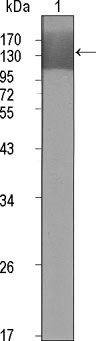
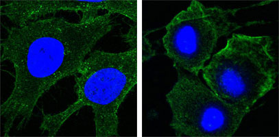
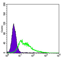
| WB, FC, ICC, E |
|---|---|
| Primary Accession | P35968 |
| Reactivity | Human |
| Host | Mouse |
| Clonality | Monoclonal |
| Clone Names | 4B4 |
| Isotype | IgG1 |
| Calculated MW | 151527 Da |
| Description | KDR has also been designated as VEFR-2 (Vascular endothelial growth factor receptor 2), CD309 (cluster of differentiation 309) and Flk1 (fetal liver kinase 1). Vascular endothelial growth factor (VEGF) is a major growth factor for endothelial cells. KDR is one of the two receptors of the VEGF. This receptor, known as kinase insert domain receptor, is a type III receptor tyrosine kinase. It functions as the main mediator of VEGF-induced endothelial proliferation, survival, migration, tubular morphogenesis and sprouting. The signalling and trafficking of this receptor are regulated by multiple factors, including Rab GTPase, P2Y purine nucleotide receptor, integrin alphaVbeta3, T-cell protein tyrosine phosphatase, etc.. Mutations of this gene are implicated in infantile capillary hemangiomas. |
| Immunogen | Purified recombinant extracellular fragment of human KDR (aa20-764) fused with hIgGFc tag expressed in HEK293 cells. |
| Formulation | Ascitic fluid containing 0.03% sodium azide. |
| Gene ID | 3791 |
|---|---|
| Other Names | Vascular endothelial growth factor receptor 2, VEGFR-2, 2.7.10.1, Fetal liver kinase 1, FLK-1, Kinase insert domain receptor, KDR, Protein-tyrosine kinase receptor flk-1, CD309, KDR, FLK1, VEGFR2 |
| Dilution | WB~~1/500 - 1/2000 FC~~1/200 - 1/400 ICC~~N/A E~~N/A |
| Storage | Maintain refrigerated at 2-8°C for up to 6 months. For long term storage store at -20°C in small aliquots to prevent freeze-thaw cycles. |
| Precautions | KDR Antibody is for research use only and not for use in diagnostic or therapeutic procedures. |
| Name | KDR (HGNC:6307) |
|---|---|
| Synonyms | FLK1, VEGFR2 |
| Function | Tyrosine-protein kinase that acts as a cell-surface receptor for VEGFA, VEGFC and VEGFD. Plays an essential role in the regulation of angiogenesis, vascular development, vascular permeability, and embryonic hematopoiesis. Promotes proliferation, survival, migration and differentiation of endothelial cells. Promotes reorganization of the actin cytoskeleton. Isoforms lacking a transmembrane domain, such as isoform 2 and isoform 3, may function as decoy receptors for VEGFA, VEGFC and/or VEGFD. Isoform 2 plays an important role as negative regulator of VEGFA- and VEGFC-mediated lymphangiogenesis by limiting the amount of free VEGFA and/or VEGFC and preventing their binding to FLT4. Modulates FLT1 and FLT4 signaling by forming heterodimers. Binding of vascular growth factors to isoform 1 leads to the activation of several signaling cascades. Activation of PLCG1 leads to the production of the cellular signaling molecules diacylglycerol and inositol 1,4,5-trisphosphate and the activation of protein kinase C. Mediates activation of MAPK1/ERK2, MAPK3/ERK1 and the MAP kinase signaling pathway, as well as of the AKT1 signaling pathway. Mediates phosphorylation of PIK3R1, the regulatory subunit of phosphatidylinositol 3-kinase, reorganization of the actin cytoskeleton and activation of PTK2/FAK1. Required for VEGFA-mediated induction of NOS2 and NOS3, leading to the production of the signaling molecule nitric oxide (NO) by endothelial cells. Phosphorylates PLCG1. Promotes phosphorylation of FYN, NCK1, NOS3, PIK3R1, PTK2/FAK1 and SRC. |
| Cellular Location | Cell junction. Endoplasmic reticulum. Cell membrane. Note=Localized with RAP1A at cell-cell junctions (By similarity). Colocalizes with ERN1 and XBP1 in the endoplasmic reticulum in endothelial cells in a vascular endothelial growth factor (VEGF)-dependent manner (PubMed:23529610). {ECO:0000250, ECO:0000269|PubMed:23529610} [Isoform 2]: Secreted. |
| Tissue Location | Detected in cornea (at protein level). Widely expressed. |
Research Areas
For Research Use Only. Not For Use In Diagnostic Procedures.
Application Protocols
Provided below are standard protocols that you may find useful for product applications.
REFERENCES
1. Blood. 2004 Aug 1;104(3):788-94. 2. FEBS Lett. 2002 Feb 13;512(1-3):107-10. 3. EMBO J. 2001 Jun 1;20(11):2768-78.
终于等到您。ABCEPTA(百远生物)抗体产品。
点击下方“我要评价 ”按钮提交您的反馈信息,您的反馈和评价是我们最宝贵的财富之一,
我们将在1-3个工作日内处理您的反馈信息。
如有疑问,联系:0512-88856768 tech-china@abcepta.com.
¥ 1,500.00
Cat# AO1324a


