


 癌症的基本特征包括细胞增殖、血管生成、迁移、凋亡逃避机制和细胞永生等。找到癌症发生过程中这些通路的关键标记物和对应的抗体用于检测至关重要。
 为您推荐一个泛素化位点预测神器——泛素化分析工具,可以为您的蛋白的泛素化位点作出预测和评分。
 细胞自噬受体图形绘图工具为你的蛋白的细胞受体结合位点作出预测和评分,识别结合到自噬通路中的蛋白是非常重要的,便于让我们理解自噬在正常生理、病理过程中的作用,如发育、细胞分化、神经退化性疾病、压力条件下、感染和癌症。
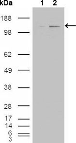
ABL2 Antibody
Purified Mouse Monoclonal Antibody
- 产品详情
- 实验流程
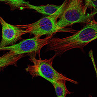
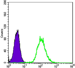
Application
| WB, ICC, E |
|---|---|
| Primary Accession | P42684 |
| Reactivity | Human, Mouse |
| Host | Mouse |
| Clonality | Monoclonal |
| Clone Names | 1H1 |
| Isotype | IgG1 |
| Calculated MW | 128343 Da |
| Description | ABL2 (ARG, Abl-related gene), together with c-Abl, forms the Abl family of mammalian non-receptor tyrosine kinases. ABL2 and c-Abl share 89%, 90 and 93% identity in their SH3, SH2 and tyrosine domain, but only 29% identity in the carboxy-terminal half. The human c-Abl and ABL2 genes are expressed ubiquitously. ABL2 had been detected predominantly in the cytoplasm, whereas c-Abl shows both cytoplasmic and nuclear localization. c-Abl is involved in two different chromosomal translocations present in human leukemias, which generate Bcr-Abl and TEL-Abl. Recently, TEL-ARG fusion transcripts have also been identified in acute myeloid leukemias (AML). The Abl family kinases may also interact with receptor tyrosine signaling pathways and regulate cellular function such as cell cycle progression, gene transcription and organization of the actin cytoskeletons in neurons. |
| Immunogen | Purified recombinant fragment of ABL2 expressed in E. Coli. |
| Formulation | Ascitic fluid containing 0.03% sodium azide. |
| Gene ID | 27 |
|---|---|
| Other Names | Abelson tyrosine-protein kinase 2, 2.7.10.2, Abelson murine leukemia viral oncogene homolog 2, Abelson-related gene protein, Tyrosine-protein kinase ARG, ABL2, ABLL, ARG |
| Dilution | WB~~1/500 - 1/2000 ICC~~N/A E~~N/A |
| Storage | Maintain refrigerated at 2-8°C for up to 6 months. For long term storage store at -20°C in small aliquots to prevent freeze-thaw cycles. |
| Precautions | ABL2 Antibody is for research use only and not for use in diagnostic or therapeutic procedures. |
| Name | ABL2 |
|---|---|
| Synonyms | ABLL, ARG |
| Function | Non-receptor tyrosine-protein kinase that plays an ABL1- overlapping role in key processes linked to cell growth and survival such as cytoskeleton remodeling in response to extracellular stimuli, cell motility and adhesion and receptor endocytosis. Coordinates actin remodeling through tyrosine phosphorylation of proteins controlling cytoskeleton dynamics like MYH10 (involved in movement); CTTN (involved in signaling); or TUBA1 and TUBB (microtubule subunits). Binds directly F-actin and regulates actin cytoskeletal structure through its F-actin- bundling activity. Involved in the regulation of cell adhesion and motility through phosphorylation of key regulators of these processes such as CRK, CRKL, DOK1 or ARHGAP35. Adhesion-dependent phosphorylation of ARHGAP35 promotes its association with RASA1, resulting in recruitment of ARHGAP35 to the cell periphery where it inhibits RHO. Phosphorylates multiple receptor tyrosine kinases like PDGFRB and other substrates which are involved in endocytosis regulation such as RIN1. In brain, may regulate neurotransmission by phosphorylating proteins at the synapse. ABL2 also acts as a regulator of multiple pathological signaling cascades during infection. Pathogens can highjack ABL2 kinase signaling to reorganize the host actin cytoskeleton for multiple purposes, like facilitating intracellular movement and host cell exit. Finally, functions as its own regulator through autocatalytic activity as well as through phosphorylation of its inhibitor, ABI1. Positively regulates chemokine-mediated T-cell migration, polarization, and homing to lymph nodes and immune-challenged tissues, potentially via activation of NEDD9/HEF1 and RAP1 (By similarity). |
| Cellular Location | Cytoplasm, cytoskeleton {ECO:0000250|UniProtKB:Q4JIM5} |
| Tissue Location | Widely expressed. |
Research Areas
For Research Use Only. Not For Use In Diagnostic Procedures.
Application Protocols
Provided below are standard protocols that you may find useful for product applications.
REFERENCES
1. Yoshimi I, Takashi I, Tsuneyuki O, et al. Blood. 2000; 95(6): 2126-2131. 2. Scheijen, B. and Griffin, J.D. Oncogene. 2002); 21:3314-33.
终于等到您。ABCEPTA(百远生物)抗体产品。
点击下方“我要评价 ”按钮提交您的反馈信息,您的反馈和评价是我们最宝贵的财富之一,
我们将在1-3个工作日内处理您的反馈信息。
如有疑问,联系:0512-88856768 tech-china@abcepta.com.
¥ 1,500.00
Cat# AO1349a


