


 癌症的基本特征包括细胞增殖、血管生成、迁移、凋亡逃避机制和细胞永生等。找到癌症发生过程中这些通路的关键标记物和对应的抗体用于检测至关重要。
 为您推荐一个泛素化位点预测神器——泛素化分析工具,可以为您的蛋白的泛素化位点作出预测和评分。
 细胞自噬受体图形绘图工具为你的蛋白的细胞受体结合位点作出预测和评分,识别结合到自噬通路中的蛋白是非常重要的,便于让我们理解自噬在正常生理、病理过程中的作用,如发育、细胞分化、神经退化性疾病、压力条件下、感染和癌症。
ATP2C1 Antibody
Purified Mouse Monoclonal Antibody
- 产品详情
- 实验流程
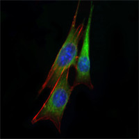
Application
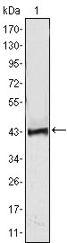
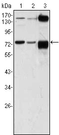
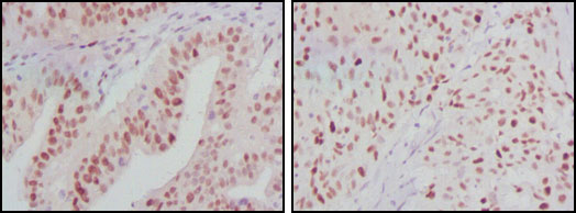
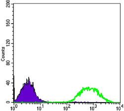
| WB, IHC, E |
|---|---|
| Primary Accession | P98194 |
| Reactivity | Human, Monkey |
| Host | Mouse |
| Clonality | Monoclonal |
| Clone Names | 4G12 |
| Isotype | IgG1 |
| Calculated MW | 100577 Da |
| Description | ATP2C1, also known as PMR1, it belongs to the family of P-type cation transport ATPases. This magnesium-dependent enzyme catalyzes the hydrolysis of ATP coupled with the transport of the calcium. The human homologue, ATP2C1 (alsodesignated SPLA in rat), also regulates the transport of calcium in the Golgicomplex and is related to other P-type ATPases family members, such as thesarco(endo)plasmic calcium ATPase (SERCA) and the plasma membrane calciumATPase (PCMA). ATP2C1 is a transmembrane protein that exists as twosplice variants, which vary by 20 amino acids. Defects in ATP2C1 cause Hailey-Hailey disease, which is an autosomal dominant disorder that is characterized by blisters and erosions of the skin. These findings provide further evidence that PMR1 plays a key role in maintaining the integrity of the epidermis by controlling intracellular calcium signaling. |
| Immunogen | Purified recombinant fragment of ATP2C1 expressed in E. Coli. |
| Formulation | Ascitic fluid containing 0.03% sodium azide. |
| Gene ID | 27032 |
|---|---|
| Other Names | Calcium-transporting ATPase type 2C member 1, ATPase 2C1, 3.6.3.8, ATP-dependent Ca(2+) pump PMR1, ATP2C1, KIAA1347, PMR1L |
| Dilution | WB~~1/500 - 1/2000 IHC~~1/500 - 1/2000 E~~N/A |
| Storage | Maintain refrigerated at 2-8°C for up to 6 months. For long term storage store at -20°C in small aliquots to prevent freeze-thaw cycles. |
| Precautions | ATP2C1 Antibody is for research use only and not for use in diagnostic or therapeutic procedures. |
| Name | ATP2C1 {ECO:0000303|PubMed:10615129, ECO:0000312|HGNC:HGNC:13211} |
|---|---|
| Function | ATP-driven pump that supplies the Golgi apparatus with Ca(2+) and Mn(2+) ions, both essential cofactors for processing and trafficking of newly synthesized proteins in the secretory pathway (PubMed:12707275, PubMed:16192278, PubMed:20439740, PubMed:21187401, PubMed:30923126). Within a catalytic cycle, acquires Ca(2+) or Mn(2+) ions on the cytoplasmic side of the membrane and delivers them to the lumenal side. The transfer of ions across the membrane is coupled to ATP hydrolysis and is associated with a transient phosphorylation that shifts the pump conformation from inward-facing to outward-facing state (PubMed:16192278, PubMed:16332677, PubMed:30923126). Plays a primary role in the maintenance of Ca(2+) homeostasis in the trans-Golgi compartment with a functional impact on Golgi and post-Golgi protein sorting as well as a structural impact on cisternae morphology (PubMed:14632183, PubMed:20439740). Responsible for loading the Golgi stores with Ca(2+) ions in keratinocytes, contributing to keratinocyte differentiation and epidermis integrity (PubMed:10615129, PubMed:14632183, PubMed:20439740). Participates in Ca(2+) and Mn(2+) ions uptake into the Golgi store of hippocampal neurons and regulates protein trafficking required for neural polarity (By similarity). May also play a role in the maintenance of Ca(2+) and Mn(2+) homeostasis and signaling in the cytosol while preventing cytotoxicity (PubMed:21187401). |
| Cellular Location | Golgi apparatus, trans-Golgi network membrane; Multi-pass membrane protein. Golgi apparatus, Golgi stack membrane; Multi-pass membrane protein. Note=During neuron differentiation, shifts from juxtanuclear Golgi position to multiple Golgi structures distributed over the neural soma with a predominance in the apical dendritic trunk {ECO:0000250|UniProtKB:Q80XR2} |
| Tissue Location | Found in most tissues except colon, thymus, spleen and leukocytes (PubMed:15831496). Expressed in keratinocytes (at protein level) (PubMed:14632183, PubMed:15831496) |
Research Areas
For Research Use Only. Not For Use In Diagnostic Procedures.
Application Protocols
Provided below are standard protocols that you may find useful for product applications.
REFERENCES
1. J Invest Dermatol. 2005 Nov;125(5):933-5. 2. J Dermatol Sci. 2006 Aug;43(2):150-1. 3. Dermatology. 2007;215(4):277-83.
终于等到您。ABCEPTA(百远生物)抗体产品。
点击下方“我要评价 ”按钮提交您的反馈信息,您的反馈和评价是我们最宝贵的财富之一,
我们将在1-3个工作日内处理您的反馈信息。
如有疑问,联系:0512-88856768 tech-china@abcepta.com.
¥ 1,500.00
Cat# AO1354a


