


 癌症的基本特征包括细胞增殖、血管生成、迁移、凋亡逃避机制和细胞永生等。找到癌症发生过程中这些通路的关键标记物和对应的抗体用于检测至关重要。
 为您推荐一个泛素化位点预测神器——泛素化分析工具,可以为您的蛋白的泛素化位点作出预测和评分。
 细胞自噬受体图形绘图工具为你的蛋白的细胞受体结合位点作出预测和评分,识别结合到自噬通路中的蛋白是非常重要的,便于让我们理解自噬在正常生理、病理过程中的作用,如发育、细胞分化、神经退化性疾病、压力条件下、感染和癌症。
ICAM1 Antibody
Purified Mouse Monoclonal Antibody
- 产品详情
- 实验流程
Application
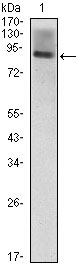
| WB, E |
|---|---|
| Primary Accession | P05362 |
| Reactivity | Human |
| Host | Mouse |
| Clonality | Monoclonal |
| Clone Names | 6G12 |
| Isotype | IgG1 |
| Calculated MW | 57825 Da |
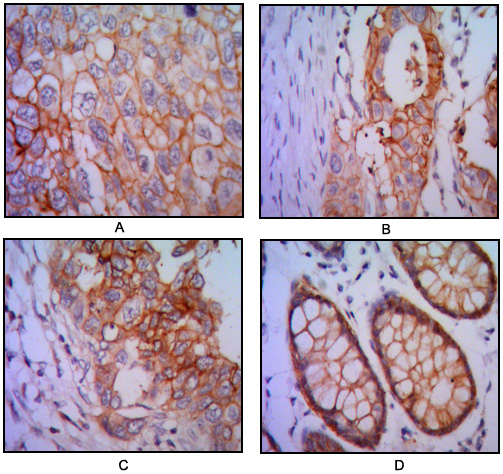
| Description | ICAM1 is a 85-110 kDa single chain type 1 integral membrane glycoprotein with an extracellular domain of five immunoglobulin superfamily repeats, a transmembrane region and a cytoplasmic domain. It shares considerable amino acid sequence homology with ICAM3 and with ICAM2. ICAM1 is expressed by activated endothelial cells. It is detected on cells of many other lineages (e.g. epithelial cells, fibroblasts, chondrocytes, B lymphocytes, T lymphocytes (low), monocytes, macrophages, dendritic cells and neutrophils), with lower levels that increase in inflammation. ICAM1 is also detected in some carcinoma and melanoma cells. Soluble ICAM1 is detectable in the plasma and is elevated in patients with various inflammatory syndromes. It is the receptor for rhinoviruses and malaria. |
| Immunogen | Purified recombinant fragment of human ICAM1(28-480aa) expressed in E. Coli. |
| Formulation | Ascitic fluid containing 0.03% sodium azide. |
| Gene ID | 3383 |
|---|---|
| Other Names | Intercellular adhesion molecule 1, ICAM-1, Major group rhinovirus receptor, CD54, ICAM1 |
| Dilution | WB~~1/500 - 1/2000 E~~N/A |
| Storage | Maintain refrigerated at 2-8°C for up to 6 months. For long term storage store at -20°C in small aliquots to prevent freeze-thaw cycles. |
| Precautions | ICAM1 Antibody is for research use only and not for use in diagnostic or therapeutic procedures. |
| Name | ICAM1 (HGNC:5344) |
|---|---|
| Function | Cell adhesion molecule that functions as a receptor ligand of the signaling receptor ITGAL:ITGB2/LFA-1 (lymphocyte-function associated (LFA) molecule 1) ensuring leukocyte cell-cell adhesion, by providing a calibrated system to namely adjust T-cell killing to the antigen stimulation strength (PubMed:3086451, PubMed:3340213, PubMed:38195629). Also functions as a ligand receptor of the signaling receptor ITGAM:ITGB2/MAC-1 ensuring adhesion between stimulated neutrophils and stimulated endothelial cells (PubMed:1980124). During leukocyte trans-endothelial migration, ICAM1 engagement promotes the assembly of endothelial apical cups through ARHGEF26/SGEF and RHOG activation (PubMed:17875742). Promotes cell aggregation in epithelial cells through interaction with MUC1 (PubMed:11173916). |
| Cellular Location | Cell membrane; Single-pass type I membrane protein |
| Tissue Location | Expressed on non-hematopoietic cells such as vascular endothelial cells, thymic epithelial cells, certain other epithelial cells, and fibroblasts, and on hematopoietic cells such as tissue macrophages, mitogen-stimulated T lymphocyte blasts, and germinal center dendritic cells in tonsils, lymph nodes, and Peyer's patches (PubMed:3086451). Expressed in low amounts on peripheral blood leukocytes (PubMed:3086451). |
Research Areas
For Research Use Only. Not For Use In Diagnostic Procedures.
Application Protocols
Provided below are standard protocols that you may find useful for product applications.
REFERENCES
1. J Neurosci Res. 1992 Feb;31(2):365-74. 2. J Cell Biol. 2002 Jun 24;157(7):1233-45. 3. J Hepatol. 2004 Mar;40(3):375-9.
终于等到您。ABCEPTA(百远生物)抗体产品。
点击下方“我要评价 ”按钮提交您的反馈信息,您的反馈和评价是我们最宝贵的财富之一,
我们将在1-3个工作日内处理您的反馈信息。
如有疑问,联系:0512-88856768 tech-china@abcepta.com.
¥ 1,500.00
Cat# AO1383a


