


 癌症的基本特征包括细胞增殖、血管生成、迁移、凋亡逃避机制和细胞永生等。找到癌症发生过程中这些通路的关键标记物和对应的抗体用于检测至关重要。
 为您推荐一个泛素化位点预测神器——泛素化分析工具,可以为您的蛋白的泛素化位点作出预测和评分。
 细胞自噬受体图形绘图工具为你的蛋白的细胞受体结合位点作出预测和评分,识别结合到自噬通路中的蛋白是非常重要的,便于让我们理解自噬在正常生理、病理过程中的作用,如发育、细胞分化、神经退化性疾病、压力条件下、感染和癌症。
EPCAM Antibody
Purified Mouse Monoclonal Antibody
- 产品详情
- 实验流程
Application
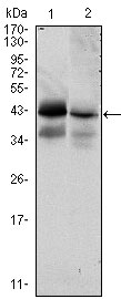
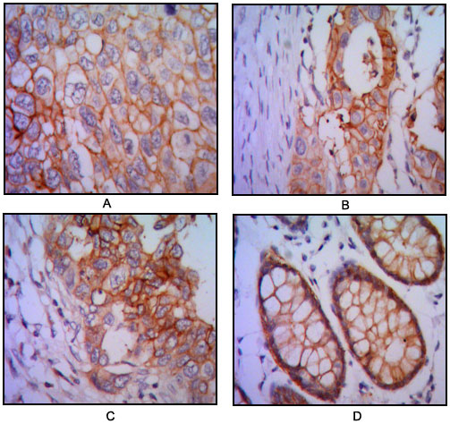
| WB, IHC, E |
|---|---|
| Primary Accession | P16422 |
| Reactivity | Human |
| Host | Mouse |
| Clonality | Monoclonal |
| Clone Names | 7E11 |
| Isotype | IgG1 |
| Calculated MW | 34932 Da |
| Description | This gene encodes a carcinoma-associated antigen and is a member of a family that includes at least two type I membrane proteins. This antigen is expressed on most normal epithelial cells and gastrointestinal carcinomas and functions as a homotypic calcium-independent cell adhesion molecule. The antigen is being used as a target for immunotherapy treatment of human carcinomas. Mutations in this gene result in congenital tufting enteropathy.Tissue specificity: This protein is expressed in almost all epithelial cell membranes but not on mesodermal or neural cell membranes. Found on the surface of adenocarcinomas.ABCAM:Epithelial Cell Adhesion Molecule (EpCAM) is a 40 kDa cell surface antigen. This antigen has been identified independently by a number of groups, and has been known by a variety of names. Several monoclonal antibodies have been raised against EpCAM, many of which have been described as tumour specific molecules on carcinomas. EpCAM is a Type 1 transmembrane glycoprotein. It is expressed on the basolateral membrane of cells by the majority of epithelial tissues, with the exception of adult squamous epithelium and some specific epithelial cell types including hepatocytes and gastric epithelial cells. EpCAM expression has been reported to be a possible marker of early malignancy, with expression being increased in tumour cells, and de novo expression being seen in dysplastic squamous epithelium.BIOLEGEND:This cell surface, glycosyl;ated 40kD protein is highly expressed in the bone marrow, colon, lung, and most normal epithelial cells and is expressed on carcinomas of gastrointestinal origin. |
| Immunogen | Purified recombinant fragment of human EPCAM expressed in E. Coli. |
| Formulation | Ascitic fluid containing 0.03% sodium azide. |
| Other Names | Epithelial cell adhesion molecule, Ep-CAM, Adenocarcinoma-associated antigen, Cell surface glycoprotein Trop-1, Epithelial cell surface antigen, Epithelial glycoprotein, EGP, Epithelial glycoprotein 314, EGP314, hEGP314, KS 1/4 antigen, KSA, Major gastrointestinal tumor-associated protein GA733-2, Tumor-associated calcium signal transducer 1, CD326, EPCAM, GA733-2, M1S2, M4S1, MIC18, TACSTD1, TROP1 |
|---|---|
| Dilution | WB~~1/500 - 1/2000 IHC~~1/200 - 1/1000 E~~N/A |
| Storage | Maintain refrigerated at 2-8°C for up to 6 months. For long term storage store at -20°C in small aliquots to prevent freeze-thaw cycles. |
| Precautions | EPCAM Antibody is for research use only and not for use in diagnostic or therapeutic procedures. |
Research Areas
For Research Use Only. Not For Use In Diagnostic Procedures.
Application Protocols
Provided below are standard protocols that you may find useful for product applications.
REFERENCES
1. Int J Oncol. 2007 Jan;30(1):171-9. 2. Int J Cancer. 2008 Jul 15;123(2):484-9.
终于等到您。ABCEPTA(百远生物)抗体产品。
点击下方“我要评价 ”按钮提交您的反馈信息,您的反馈和评价是我们最宝贵的财富之一,
我们将在1-3个工作日内处理您的反馈信息。
如有疑问,联系:0512-88856768 tech-china@abcepta.com.
¥ 1,500.00
Cat# AO1402a


