


 癌症的基本特征包括细胞增殖、血管生成、迁移、凋亡逃避机制和细胞永生等。找到癌症发生过程中这些通路的关键标记物和对应的抗体用于检测至关重要。
 为您推荐一个泛素化位点预测神器——泛素化分析工具,可以为您的蛋白的泛素化位点作出预测和评分。
 细胞自噬受体图形绘图工具为你的蛋白的细胞受体结合位点作出预测和评分,识别结合到自噬通路中的蛋白是非常重要的,便于让我们理解自噬在正常生理、病理过程中的作用,如发育、细胞分化、神经退化性疾病、压力条件下、感染和癌症。
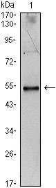
IL34 Antibody
Purified Mouse Monoclonal Antibody
- 产品详情
- 实验流程
Application
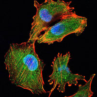
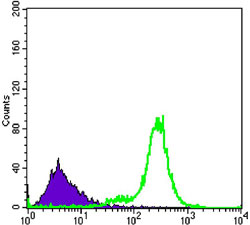
| WB, FC, ICC, E |
|---|---|
| Primary Accession | Q6ZMJ4 |
| Reactivity | Human |
| Host | Mouse |
| Clonality | Monoclonal |
| Clone Names | 1D12 |
| Isotype | IgG1 |
| Calculated MW | 27482 Da |
| Description | Cytokine that promotes the differentiation and viability of monocytes and macrophages. Stimulates phosphorylation of MAPK1/ERK2 AND MAPK3/ERK1. Ligand for colony-stimulating factor-1 receptor CSF1R.Tissue specificity: Detected in the sinusoidal epithelium in the red pulp of spleen (at protein level). Predominantly expressed in spleen. Also detected in a range of other tissues including heart, brain, lung, liver, kidney, thymus, testis, ovary, small intestine, prostate and colon. |
| Immunogen | Purified recombinant fragment of human IL34 expressed in E. Coli. |
| Formulation | Ascitic fluid containing 0.03% sodium azide. |
| Gene ID | 146433 |
|---|---|
| Other Names | Interleukin-34, IL-34, IL34, C16orf77 |
| Dilution | WB~~1/500 - 1/2000 FC~~1/200 - 1/400 ICC~~N/A E~~N/A |
| Storage | Maintain refrigerated at 2-8°C for up to 6 months. For long term storage store at -20°C in small aliquots to prevent freeze-thaw cycles. |
| Precautions | IL34 Antibody is for research use only and not for use in diagnostic or therapeutic procedures. |
| Name | IL34 |
|---|---|
| Synonyms | C16orf77 |
| Function | Cytokine that promotes the proliferation, survival and differentiation of monocytes and macrophages. Promotes the release of pro-inflammatory chemokines, and thereby plays an important role in innate immunity and in inflammatory processes. Plays an important role in the regulation of osteoclast proliferation and differentiation, and in the regulation of bone resorption. Signaling via CSF1R and its downstream effectors stimulates phosphorylation of MAPK1/ERK2 AND MAPK3/ERK1. |
| Cellular Location | Secreted. |
| Tissue Location | Detected in the sinusoidal epithelium in the red pulp of spleen (at protein level). Predominantly expressed in spleen Also detected in a range of other tissues including heart, brain, lung, liver, kidney, thymus, testis, ovary, small intestine, prostate and colon. |
Research Areas
For Research Use Only. Not For Use In Diagnostic Procedures.
Application Protocols
Provided below are standard protocols that you may find useful for product applications.
REFERENCES
1. Strausberg RL, et al. Proc Natl Acad Sci U S A, 2002 Dec 24. 2. Lim J, et al. Cell, 2006 May 19. 3. Lin H, et al. Science, 2008 May 9.
终于等到您。ABCEPTA(百远生物)抗体产品。
点击下方“我要评价 ”按钮提交您的反馈信息,您的反馈和评价是我们最宝贵的财富之一,
我们将在1-3个工作日内处理您的反馈信息。
如有疑问,联系:0512-88856768 tech-china@abcepta.com.


