


 癌症的基本特征包括细胞增殖、血管生成、迁移、凋亡逃避机制和细胞永生等。找到癌症发生过程中这些通路的关键标记物和对应的抗体用于检测至关重要。
 为您推荐一个泛素化位点预测神器——泛素化分析工具,可以为您的蛋白的泛素化位点作出预测和评分。
 细胞自噬受体图形绘图工具为你的蛋白的细胞受体结合位点作出预测和评分,识别结合到自噬通路中的蛋白是非常重要的,便于让我们理解自噬在正常生理、病理过程中的作用,如发育、细胞分化、神经退化性疾病、压力条件下、感染和癌症。
HK2 Antibody
Purified Mouse Monoclonal Antibody
- 产品详情
- 实验流程
Application
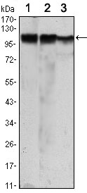
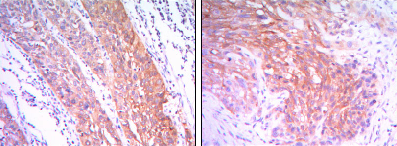
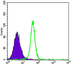
| WB, IHC, FC, E |
|---|---|
| Primary Accession | P52789 |
| Reactivity | Human |
| Host | Mouse |
| Clonality | Monoclonal |
| Clone Names | 3D3 |
| Isotype | IgG1 |
| Calculated MW | 102380 Da |
| Description | The hexokinases utilize Mg-ATP as a phosphoryl donor to catalyze the first step of intracellular glucose metabolism, the conversion of glucose to glucose- 6-phosphate. Four hexokinase isoenzymes have been identified, including hexokinase I (HXK I), hexokinase II (HXK II), hexokinase III (HXK III) and hexokinase IV (HXK IV, also designated glucokinase or GCK). Hexokinases I-III each contain an N-terminal cluster of hydrophobic amino acids. Glucokinase lacks the N-terminal hydrophobic cluster. The hydrophobic cluster is thought to be necessary for membrane binding. This is substantiated by the finding that glucokinase has lower affinity for glucose than do the other hexokinases .Hexokinase 2 is the predominant hexokinase isozyme expressed in insulin-responsive tissues such as skeletal muscle. Expression of this gene is insulin-responsive, and studies in rat suggest that it is involved in the increased rate of glycolysis seen in rapidly growing cancer cells. |
| Immunogen | Purified recombinant fragment of human HK2 expressed in E. Coli. |
| Formulation | Ascitic fluid containing 0.03% sodium azide. |
| Gene ID | 3099 |
|---|---|
| Other Names | Hexokinase-2, 2.7.1.1, Hexokinase type II, HK II, Muscle form hexokinase, HK2 |
| Dilution | WB~~1/500 - 1/2000 IHC~~1/200 - 1/1000 FC~~1/200 - 1/400 E~~N/A |
| Storage | Maintain refrigerated at 2-8°C for up to 6 months. For long term storage store at -20°C in small aliquots to prevent freeze-thaw cycles. |
| Precautions | HK2 Antibody is for research use only and not for use in diagnostic or therapeutic procedures. |
| Name | HK2 (HGNC:4923) |
|---|---|
| Function | Catalyzes the phosphorylation of hexose, such as D-glucose and D-fructose, to hexose 6-phosphate (D-glucose 6-phosphate and D- fructose 6-phosphate, respectively) (PubMed:23185017, PubMed:26985301, PubMed:29298880). Mediates the initial step of glycolysis by catalyzing phosphorylation of D-glucose to D-glucose 6-phosphate (PubMed:29298880). Plays a key role in maintaining the integrity of the outer mitochondrial membrane by preventing the release of apoptogenic molecules from the intermembrane space and subsequent apoptosis (PubMed:18350175). |
| Cellular Location | Mitochondrion outer membrane; Peripheral membrane protein. Cytoplasm, cytosol Note=The mitochondrial-binding peptide (MBP) region promotes association with the mitochondrial outer membrane (PubMed:29298880) The interaction with the mitochondrial outer membrane via the mitochondrial-binding peptide (MBP) region promotes higher stability of the protein (PubMed:29298880). Release from the mitochondrial outer membrane into the cytosol induces permeability transition pore (PTP) opening and apoptosis (PubMed:18350175). |
| Tissue Location | Predominant hexokinase isozyme expressed in insulin-responsive tissues such as skeletal muscle |
Research Areas
For Research Use Only. Not For Use In Diagnostic Procedures.
Application Protocols
Provided below are standard protocols that you may find useful for product applications.
REFERENCES
1. Cell. 2006 May 19;125(4):801-14. 2. Cancer Sci. 2008 Feb;99(2):260-6. 3. Med Oncol. 2009;26(3):303-8.
终于等到您。ABCEPTA(百远生物)抗体产品。
点击下方“我要评价 ”按钮提交您的反馈信息,您的反馈和评价是我们最宝贵的财富之一,
我们将在1-3个工作日内处理您的反馈信息。
如有疑问,联系:0512-88856768 tech-china@abcepta.com.
¥ 1,500.00
Cat# AO1441a


