


 癌症的基本特征包括细胞增殖、血管生成、迁移、凋亡逃避机制和细胞永生等。找到癌症发生过程中这些通路的关键标记物和对应的抗体用于检测至关重要。
 为您推荐一个泛素化位点预测神器——泛素化分析工具,可以为您的蛋白的泛素化位点作出预测和评分。
 细胞自噬受体图形绘图工具为你的蛋白的细胞受体结合位点作出预测和评分,识别结合到自噬通路中的蛋白是非常重要的,便于让我们理解自噬在正常生理、病理过程中的作用,如发育、细胞分化、神经退化性疾病、压力条件下、感染和癌症。
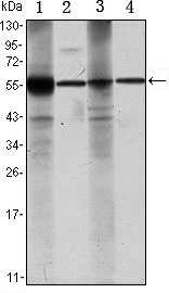
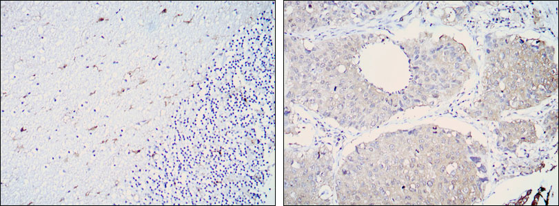
ALDH1A1 Antibody
Purified Mouse Monoclonal Antibody
- 产品详情
- 实验流程
Application
| WB, IHC, E |
|---|---|
| Primary Accession | P00352 |
| Reactivity | Human |
| Host | Mouse |
| Clonality | Monoclonal |
| Clone Names | 5A11 |
| Isotype | IgG1 |
| Calculated MW | 54862 Da |
| Description | ALDH1A1 is an aldehyde dehydrogenase able to oxidize a wide variety of aliphatic aldehydes (retinaldehyde, acetaldehyde, etc.) to the corresponding carboxylic acids (retinoic acid, acetic acid, etc.). ALDH1A1 (also known as RALDH1, ALDH1, or AHD2) is highly expressed in the dorsal retina, ventral midbrain (dopaminergic neurons), and hematopoietic stem cells. ALDH1A1 is involved in retinoic acid (RA) synthesis during vertebrate embryogenesis. ALDH1A1 is first detected at E9.0-E10.5 in cranial tissues (ventral mesencephalon, dorsal retina, thymic primordia, optic vesicles) and in the mesonephros. ALDH1A1 is also of interest in Parkinson's Disease (PD) being expressed in the the A9 dopaminergic (DA) neuronal group projecting to the dorsal striatum; this is the most vulnerable site in PD (Chung et al, 2005). ALDH1A1 protein is a known mesencephalic dopaminergic marker. ALDH1A1 is a cytosolic enzyme that preferentially oxidizes retinaldehyde to retinoic acid .ALDH1A1 is expressed in the epithelium of many organs, including brain, liver, testis, eye lens and cornea .ALDH1A1 is highly expressed in brain dopaminergic neurons, where it produces the retinoic acid required for their differentiation and development .The retinoic acid produced by ALDH1A1 is also important for the differentiation of hematopoietic stem cells. |
| Immunogen | Purified recombinant fragment of human ALDH1A1 expressed in E. Coli. |
| Formulation | Ascitic fluid containing 0.03% sodium azide. |
| Other Names | Retinal dehydrogenase 1, RALDH 1, RalDH1, 1.2.1.36, ALDH-E1, ALHDII, Aldehyde dehydrogenase family 1 member A1, Aldehyde dehydrogenase, cytosolic, ALDH1A1, ALDC, ALDH1, PUMB1 |
|---|---|
| Dilution | WB~~1/500 - 1/2000 IHC~~1/200 - 1/1000 E~~N/A |
| Storage | Maintain refrigerated at 2-8°C for up to 6 months. For long term storage store at -20°C in small aliquots to prevent freeze-thaw cycles. |
| Precautions | ALDH1A1 Antibody is for research use only and not for use in diagnostic or therapeutic procedures. |
Research Areas
For Research Use Only. Not For Use In Diagnostic Procedures.
Application Protocols
Provided below are standard protocols that you may find useful for product applications.
REFERENCES
1. J Hum Genet. 2009 Jun;54(6):317-23. 2. J Hum Genet. 2009 Oct;54(10):564-71. 3. J Neurosci Res. 2010 Feb 15;88(3):686-94.
终于等到您。ABCEPTA(百远生物)抗体产品。
点击下方“我要评价 ”按钮提交您的反馈信息,您的反馈和评价是我们最宝贵的财富之一,
我们将在1-3个工作日内处理您的反馈信息。
如有疑问,联系:0512-88856768 tech-china@abcepta.com.
¥ 1,500.00
Cat# AO1454a


