


 癌症的基本特征包括细胞增殖、血管生成、迁移、凋亡逃避机制和细胞永生等。找到癌症发生过程中这些通路的关键标记物和对应的抗体用于检测至关重要。
 为您推荐一个泛素化位点预测神器——泛素化分析工具,可以为您的蛋白的泛素化位点作出预测和评分。
 细胞自噬受体图形绘图工具为你的蛋白的细胞受体结合位点作出预测和评分,识别结合到自噬通路中的蛋白是非常重要的,便于让我们理解自噬在正常生理、病理过程中的作用,如发育、细胞分化、神经退化性疾病、压力条件下、感染和癌症。
GFAP Antibody
Purified Mouse Monoclonal Antibody
- 产品详情
- 实验流程
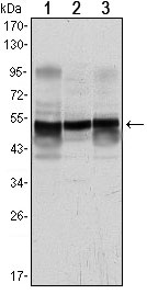
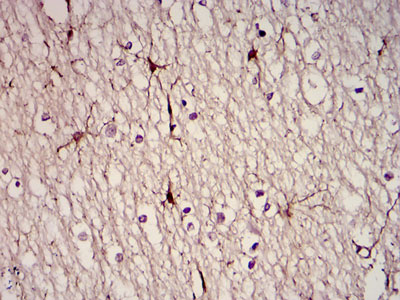
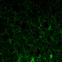
Application
| WB, IHC, ICC, E |
|---|---|
| Primary Accession | P14136 |
| Reactivity | Human |
| Host | Mouse |
| Clonality | Monoclonal |
| Clone Names | 6A6 |
| Isotype | IgG1 |
| Calculated MW | 49880 Da |
| Description | GFAP, a class-III intermediate filament, is a cell-specific marker that, during the development of the central nervous system, distinguishes astrocytes from other glial cells.Tissue specificity: Expressed in cells lacking fibronectin.ABCAM:It is heavily, and specifically, expressed in astrocytes and certain other astroglia in the central nervous system, in satellite cells in peripheral ganglia, and in non myelinating Schwann cells in peripheral nerves.In addition many types of brain tumor, presumably derived from astrocytic cells, heavily express GFAP. GFAP is also found in the lens epithelium, Kupffer cells of the liver, in some cells in salivary tumors and has been reported in erythrocytes. |
| Immunogen | Purified recombinant fragment of human GFAP expressed in E. Coli. |
| Formulation | Ascitic fluid containing 0.03% sodium azide. |
| Gene ID | 2670 |
|---|---|
| Other Names | Glial fibrillary acidic protein, GFAP, GFAP |
| Dilution | WB~~1/500 - 1/2000 IHC~~1/200 - 1/1000 ICC~~N/A E~~N/A |
| Storage | Maintain refrigerated at 2-8°C for up to 6 months. For long term storage store at -20°C in small aliquots to prevent freeze-thaw cycles. |
| Precautions | GFAP Antibody is for research use only and not for use in diagnostic or therapeutic procedures. |
| Name | GFAP |
|---|---|
| Function | GFAP, a class-III intermediate filament, is a cell-specific marker that, during the development of the central nervous system, distinguishes astrocytes from other glial cells. |
| Cellular Location | Cytoplasm. Note=Associated with intermediate filaments |
| Tissue Location | Expressed in cells lacking fibronectin. |
Research Areas
For Research Use Only. Not For Use In Diagnostic Procedures.
Application Protocols
Provided below are standard protocols that you may find useful for product applications.
REFERENCES
1. Acta Neuropathol. 2009 Jun;117(6):667-75. 2. Schizophr Res. 2009 Jul;112(1-3):54-64.
终于等到您。ABCEPTA(百远生物)抗体产品。
点击下方“我要评价 ”按钮提交您的反馈信息,您的反馈和评价是我们最宝贵的财富之一,
我们将在1-3个工作日内处理您的反馈信息。
如有疑问,联系:0512-88856768 tech-china@abcepta.com.
¥ 1,500.00
Cat# AO1460a


