


 癌症的基本特征包括细胞增殖、血管生成、迁移、凋亡逃避机制和细胞永生等。找到癌症发生过程中这些通路的关键标记物和对应的抗体用于检测至关重要。
 为您推荐一个泛素化位点预测神器——泛素化分析工具,可以为您的蛋白的泛素化位点作出预测和评分。
 细胞自噬受体图形绘图工具为你的蛋白的细胞受体结合位点作出预测和评分,识别结合到自噬通路中的蛋白是非常重要的,便于让我们理解自噬在正常生理、病理过程中的作用,如发育、细胞分化、神经退化性疾病、压力条件下、感染和癌症。
CDH1 Antibody
Purified Mouse Monoclonal Antibody
- 产品详情
- 实验流程
Application
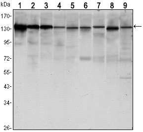
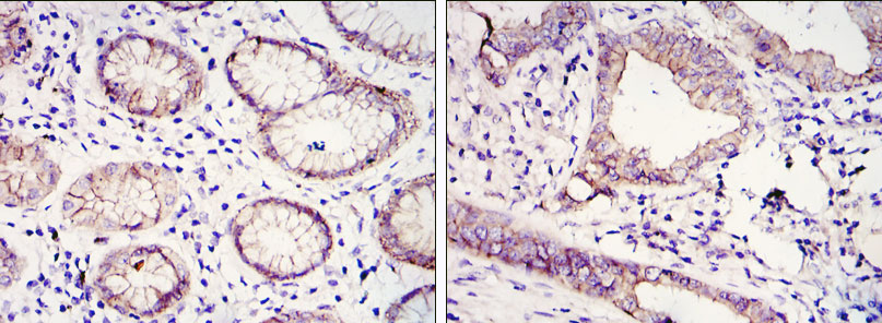
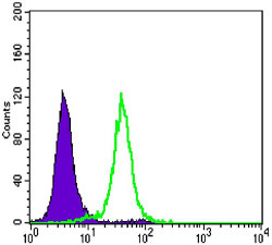
| WB, IHC, FC, E |
|---|---|
| Primary Accession | P12830 |
| Reactivity | Human, Mouse, Monkey |
| Host | Mouse |
| Clonality | Monoclonal |
| Clone Names | 7H12 |
| Isotype | IgG1 |
| Calculated MW | 97456 Da |
| Description | E-Cadherin is a 120 kDa transmembrane glycoprotein that is localized in the adherens junctions of epithelial cells. There, it interacts with the cytoskeleton through the associated cytoplasmic catenin proteins. In addition to being a calcium-dependent adhesion molecule, E-Cadherin is also a critical regulator of epithelial junction formation. Its association with catenins is necessary for cell-cell adhesion. These E-cadherin/catenin complexes associate with corical actin bundles at both the zonula adherens and the lateral adhesion plaques. Tyrosine phosphorylation can disrupt these complexes, leading to changes in cell adhesion properties. E-Cadherin expression is often down-regulated in highly invasive, poorly differentiated carcinomas. Increased expression of E-Cadherin in these cells reduces invasiveness. Thus, loss of expression or function of E-Cadherin appears to be an important step in tumorigenic progression.Tissue specificity: Non-neural epithelial tissues. |
| Immunogen | Purified recombinant fragment of human CDH1 expressed in E. Coli. |
| Formulation | Ascitic fluid containing 0.03% sodium azide. |
| Other Names | Cadherin-1, CAM 120/80, Epithelial cadherin, E-cadherin, Uvomorulin, CD324, E-Cad/CTF1, E-Cad/CTF2, E-Cad/CTF3, CDH1, CDHE, UVO |
|---|---|
| Dilution | WB~~1/500 - 1/2000 IHC~~1/200 - 1/1000 FC~~1/200 - 1/400 E~~N/A |
| Storage | Maintain refrigerated at 2-8°C for up to 6 months. For long term storage store at -20°C in small aliquots to prevent freeze-thaw cycles. |
| Precautions | CDH1 Antibody is for research use only and not for use in diagnostic or therapeutic procedures. |
Research Areas
For Research Use Only. Not For Use In Diagnostic Procedures.
Application Protocols
Provided below are standard protocols that you may find useful for product applications.
REFERENCES
1. Nat Genet. 2009 Dec;41(12):1330-4. 2. Zhonghua Zhong Liu Za Zhi. 2009 Jul;31(7):515-9. 3. J Biol Chem. 2010 Feb 26;285(9):6658-69.
终于等到您。ABCEPTA(百远生物)抗体产品。
点击下方“我要评价 ”按钮提交您的反馈信息,您的反馈和评价是我们最宝贵的财富之一,
我们将在1-3个工作日内处理您的反馈信息。
如有疑问,联系:0512-88856768 tech-china@abcepta.com.
¥ 1,520.00
Cat# AO1473a


