


 癌症的基本特征包括细胞增殖、血管生成、迁移、凋亡逃避机制和细胞永生等。找到癌症发生过程中这些通路的关键标记物和对应的抗体用于检测至关重要。
 为您推荐一个泛素化位点预测神器——泛素化分析工具,可以为您的蛋白的泛素化位点作出预测和评分。
 细胞自噬受体图形绘图工具为你的蛋白的细胞受体结合位点作出预测和评分,识别结合到自噬通路中的蛋白是非常重要的,便于让我们理解自噬在正常生理、病理过程中的作用,如发育、细胞分化、神经退化性疾病、压力条件下、感染和癌症。
C-CBL Antibody
Purified Mouse Monoclonal Antibody
- 产品详情
- 实验流程
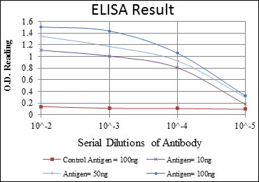
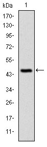
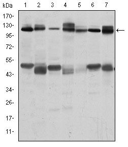
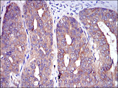
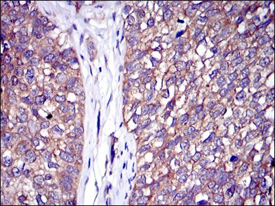
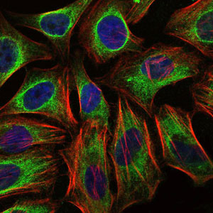
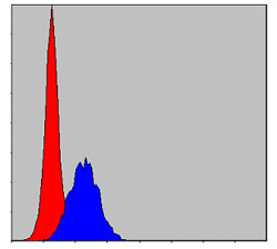
Application
| WB, IHC, FC, ICC, E |
|---|---|
| Primary Accession | P22681 |
| Reactivity | Human, Mouse, Rat |
| Host | Mouse |
| Clonality | Monoclonal |
| Clone Names | 3B12 |
| Isotype | IgG1 |
| Calculated MW | 99633 Da |
| Description | The cbl oncogene was first identified as part of a transforming retrovirus which induces mouse pre-B and pro-B cell lymphomas. As an adaptor protein for receptor protein-tyrosine kinases, it positively regulates receptor protein-tyrosine kinase ubiquitination in a manner dependent upon its variant SH2 and RING finger domains. Ubiquitination of receptor protein-tyrosine kinases terminates signaling by marking active receptors for degradation. |
| Immunogen | Purified recombinant fragment of human C-CBL expressed in E. Coli. |
| Formulation | Ascitic fluid containing 0.03% sodium azide. |
| Gene ID | 867 |
|---|---|
| Other Names | E3 ubiquitin-protein ligase CBL, 6.3.2.-, Casitas B-lineage lymphoma proto-oncogene, Proto-oncogene c-Cbl, RING finger protein 55, Signal transduction protein CBL, CBL, CBL2, RNF55 |
| Dilution | WB~~1/500 - 1/2000 IHC~~1/200 - 1/1000 FC~~1/200 - 1/400 ICC~~N/A E~~1/10000 |
| Storage | Maintain refrigerated at 2-8°C for up to 6 months. For long term storage store at -20°C in small aliquots to prevent freeze-thaw cycles. |
| Precautions | C-CBL Antibody is for research use only and not for use in diagnostic or therapeutic procedures. |
| Name | CBL |
|---|---|
| Synonyms | CBL2, RNF55 |
| Function | E3 ubiquitin-protein ligase that acts as a negative regulator of many signaling pathways by mediating ubiquitination of cell surface receptors (PubMed:10514377, PubMed:11896602, PubMed:14661060, PubMed:14739300, PubMed:15190072, PubMed:17509076, PubMed:18374639, PubMed:19689429, PubMed:21596750, PubMed:28381567). Accepts ubiquitin from specific E2 ubiquitin-conjugating enzymes, and then transfers it to substrates promoting their degradation by the proteasome (PubMed:10514377, PubMed:14661060, PubMed:14739300, PubMed:17094949, PubMed:17509076, PubMed:17974561). Recognizes activated receptor tyrosine kinases, including KIT, FLT1, FGFR1, FGFR2, PDGFRA, PDGFRB, CSF1R, EPHA8 and KDR and mediates their ubiquitination to terminate signaling (PubMed:15190072, PubMed:18374639, PubMed:21596750). Recognizes membrane-bound HCK, SRC and other kinases of the SRC family and mediates their ubiquitination and degradation (PubMed:11896602). Ubiquitinates EGFR and SPRY2 (PubMed:17094949, PubMed:17974561). Ubiquitinates NECTIN1 following association between NECTIN1 and herpes simplex virus 1/HHV-1 envelope glycoprotein D, leading to NECTIN1 removal from cell surface (PubMed:28381567). Participates in signal transduction in hematopoietic cells. Plays an important role in the regulation of osteoblast differentiation and apoptosis (PubMed:15190072, PubMed:18374639). Essential for osteoclastic bone resorption (PubMed:14739300). The 'Tyr-731' phosphorylated form induces the activation and recruitment of phosphatidylinositol 3-kinase to the cell membrane in a signaling pathway that is critical for osteoclast function (PubMed:14739300). May be functionally coupled with the E2 ubiquitin-protein ligase UB2D3. In association with CBLB, required for proper feedback inhibition of ciliary platelet-derived growth factor receptor-alpha (PDGFRA) signaling pathway via ubiquitination and internalization of PDGFRA (By similarity). |
| Cellular Location | Cytoplasm. Cell membrane. Cell projection, cilium. Golgi apparatus. Note=Colocalizes with FGFR2 in lipid rafts at the cell membrane |
Research Areas
For Research Use Only. Not For Use In Diagnostic Procedures.
Application Protocols
Provided below are standard protocols that you may find useful for product applications.
REFERENCES
1. Blood. 2009 Aug 27;114(9):1859-63. 2. Cell Res. 2009 Aug;19(8):950-61. 3. Nature. 2009 Aug 13;460(7257):904-8.
终于等到您。ABCEPTA(百远生物)抗体产品。
点击下方“我要评价 ”按钮提交您的反馈信息,您的反馈和评价是我们最宝贵的财富之一,
我们将在1-3个工作日内处理您的反馈信息。
如有疑问,联系:0512-88856768 tech-china@abcepta.com.


