


 癌症的基本特征包括细胞增殖、血管生成、迁移、凋亡逃避机制和细胞永生等。找到癌症发生过程中这些通路的关键标记物和对应的抗体用于检测至关重要。
 为您推荐一个泛素化位点预测神器——泛素化分析工具,可以为您的蛋白的泛素化位点作出预测和评分。
 细胞自噬受体图形绘图工具为你的蛋白的细胞受体结合位点作出预测和评分,识别结合到自噬通路中的蛋白是非常重要的,便于让我们理解自噬在正常生理、病理过程中的作用,如发育、细胞分化、神经退化性疾病、压力条件下、感染和癌症。
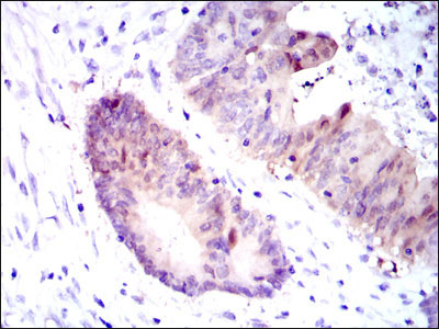
ASS1 Antibody
Purified Mouse Monoclonal Antibody
- 产品详情
- 实验流程
Application
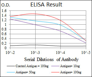
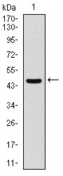
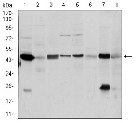
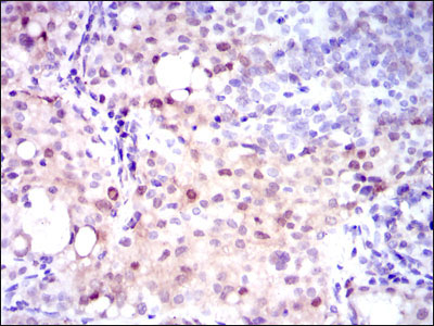
| WB, IHC, E |
|---|---|
| Primary Accession | P00966 |
| Reactivity | Human, Mouse, Monkey |
| Host | Mouse |
| Clonality | Monoclonal |
| Clone Names | 2C10 |
| Isotype | IgG1 |
| Calculated MW | 46530 Da |
| Description | The protein encoded by this gene catalyzes the penultimate step of the arginine biosynthetic pathway. There are approximately 10 to 14 copies of this gene including the pseudogenes scattered across the human genome, among which the one located on chromosome 9 appears to be the only functional gene for argininosuccinate synthetase. Mutations in the chromosome 9 copy of ASS cause citrullinemia. Two transcript variants encoding the same protein have been found for this gene. |
| Immunogen | Purified recombinant fragment of human ASS1 expressed in E. Coli. |
| Formulation | Purified antibody in PBS with 0.05% sodium azide |
| Gene ID | 445 |
|---|---|
| Other Names | Argininosuccinate synthase, 6.3.4.5, Citrulline--aspartate ligase, ASS1, ASS |
| Dilution | WB~~1/500 - 1/2000 IHC~~1/200 - 1/1000 E~~1/10000 |
| Storage | Maintain refrigerated at 2-8°C for up to 6 months. For long term storage store at -20°C in small aliquots to prevent freeze-thaw cycles. |
| Precautions | ASS1 Antibody is for research use only and not for use in diagnostic or therapeutic procedures. |
| Name | ASS1 (HGNC:758) |
|---|---|
| Function | One of the enzymes of the urea cycle, the metabolic pathway transforming neurotoxic amonia produced by protein catabolism into inocuous urea in the liver of ureotelic animals. Catalyzes the formation of arginosuccinate from aspartate, citrulline and ATP and together with ASL it is responsible for the biosynthesis of arginine in most body tissues. |
| Cellular Location | Cytoplasm, cytosol |
| Tissue Location | Expressed in adult liver. |
Research Areas
For Research Use Only. Not For Use In Diagnostic Procedures.
Application Protocols
Provided below are standard protocols that you may find useful for product applications.
REFERENCES
1. Int J Cancer. 2009 Sep 15;125(6):1454-63. 2. Clin Biochem. 2009 Jul;42(10-11):1166-8.
终于等到您。ABCEPTA(百远生物)抗体产品。
点击下方“我要评价 ”按钮提交您的反馈信息,您的反馈和评价是我们最宝贵的财富之一,
我们将在1-3个工作日内处理您的反馈信息。
如有疑问,联系:0512-88856768 tech-china@abcepta.com.
¥ 1,500.00
Cat# AO1671a


