


 癌症的基本特征包括细胞增殖、血管生成、迁移、凋亡逃避机制和细胞永生等。找到癌症发生过程中这些通路的关键标记物和对应的抗体用于检测至关重要。
 为您推荐一个泛素化位点预测神器——泛素化分析工具,可以为您的蛋白的泛素化位点作出预测和评分。
 细胞自噬受体图形绘图工具为你的蛋白的细胞受体结合位点作出预测和评分,识别结合到自噬通路中的蛋白是非常重要的,便于让我们理解自噬在正常生理、病理过程中的作用,如发育、细胞分化、神经退化性疾病、压力条件下、感染和癌症。
NOS2 Antibody
Purified Mouse Monoclonal Antibody
- 产品详情
- 实验流程
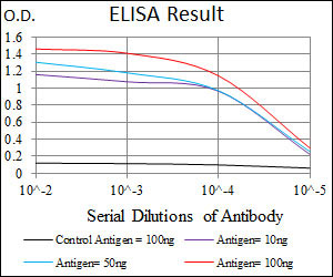
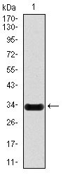
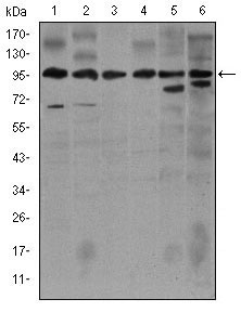
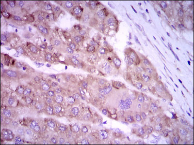
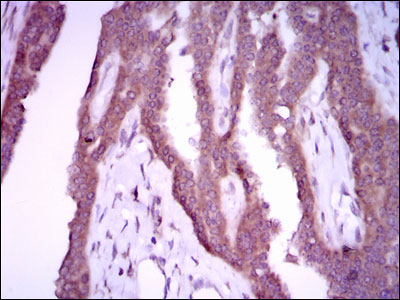
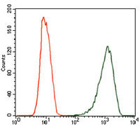
Application
| WB, IHC, FC, E |
|---|---|
| Primary Accession | P35228 |
| Reactivity | Human, Mouse |
| Host | Mouse |
| Clonality | Monoclonal |
| Clone Names | 4E5 |
| Isotype | IgG1 |
| Calculated MW | 131117 Da |
| Description | Nitric oxide is a reactive free radical which acts as a biologic mediator in several processes, including neurotransmission and antimicrobial and antitumoral activities. This gene encodes a nitric oxide synthase which is expressed in liver and is inducible by a combination of lipopolysaccharide and certain cytokines. Three related pseudogenes are located within the Smith-Magenis syndrome region on chromosome 17. |
| Immunogen | Purified recombinant fragment of human NOS2 expressed in E. Coli. |
| Formulation | Purified antibody in PBS with 0.05% sodium azide |
| Gene ID | 4843 |
|---|---|
| Other Names | Nitric oxide synthase, inducible, 1.14.13.39, Hepatocyte NOS, HEP-NOS, Inducible NO synthase, Inducible NOS, iNOS, NOS type II, Peptidyl-cysteine S-nitrosylase NOS2, NOS2, NOS2A |
| Dilution | WB~~1/500 - 1/2000 IHC~~1/200 - 1/1000 FC~~1/200 - 1/400 E~~1/10000 |
| Storage | Maintain refrigerated at 2-8°C for up to 6 months. For long term storage store at -20°C in small aliquots to prevent freeze-thaw cycles. |
| Precautions | NOS2 Antibody is for research use only and not for use in diagnostic or therapeutic procedures. |
| Name | NOS2 (HGNC:7873) |
|---|---|
| Synonyms | NOS2A |
| Function | Produces nitric oxide (NO) which is a messenger molecule with diverse functions throughout the body (PubMed:7504305, PubMed:7531687, PubMed:7544004, PubMed:7682706). In macrophages, NO mediates tumoricidal and bactericidal actions. Also has nitrosylase activity and mediates cysteine S-nitrosylation of cytoplasmic target proteins such PTGS2/COX2 (By similarity). As component of the iNOS-S100A8/9 transnitrosylase complex involved in the selective inflammatory stimulus-dependent S-nitrosylation of GAPDH on 'Cys-247' implicated in regulation of the GAIT complex activity and probably multiple targets including ANXA5, EZR, MSN and VIM (PubMed:25417112). Involved in inflammation, enhances the synthesis of pro-inflammatory mediators such as IL6 and IL8 (PubMed:19688109). |
| Cellular Location | Cytoplasm, cytosol. Note=Localizes as discrete foci scattered throughout the cytosol and in the presence of SPSB1 and SPSB4, exhibits a more diffuse cytosolic localization. |
| Tissue Location | Expressed in the liver, retina, bone cells and airway epithelial cells of the lung. Not expressed in the platelets Expressed in chondrocytes (PubMed:7504305) |
Research Areas
For Research Use Only. Not For Use In Diagnostic Procedures.
Application Protocols
Provided below are standard protocols that you may find useful for product applications.
REFERENCES
1. Pediatr Allergy Immunol. 2010 Dec;21(8):1151-6. 2. J Biol Chem. 2010 Dec 31;285(53):41422-31.
终于等到您。ABCEPTA(百远生物)抗体产品。
点击下方“我要评价 ”按钮提交您的反馈信息,您的反馈和评价是我们最宝贵的财富之一,
我们将在1-3个工作日内处理您的反馈信息。
如有疑问,联系:0512-88856768 tech-china@abcepta.com.
¥ 1,500.00
Cat# AO1718a


