


 癌症的基本特征包括细胞增殖、血管生成、迁移、凋亡逃避机制和细胞永生等。找到癌症发生过程中这些通路的关键标记物和对应的抗体用于检测至关重要。
 为您推荐一个泛素化位点预测神器——泛素化分析工具,可以为您的蛋白的泛素化位点作出预测和评分。
 细胞自噬受体图形绘图工具为你的蛋白的细胞受体结合位点作出预测和评分,识别结合到自噬通路中的蛋白是非常重要的,便于让我们理解自噬在正常生理、病理过程中的作用,如发育、细胞分化、神经退化性疾病、压力条件下、感染和癌症。
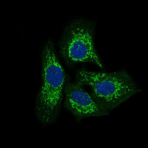
TWF1 Antibody
Purified Mouse Monoclonal Antibody


- 产品详情
- 实验流程
Application
| WB, IHC, FC, ICC, E |
|---|---|
| Primary Accession | Q12792 |
| Reactivity | Human |
| Host | Mouse |
| Clonality | Monoclonal |
| Clone Names | 7C11 |
| Isotype | IgG1 |
| Calculated MW | 40283 Da |
| Description | This gene encodes twinfilin, an actin monomer-binding protein conserved from yeast to mammals. Studies of the mouse counterpart suggest that this protein may be an actin monomer-binding protein, and its localization to cortical G-actin-rich structures may be regulated by the small GTPase RAC1. |
| Immunogen | Purified recombinant fragment of human TWF1 (AA: 335-384 ) expressed in E. Coli. |
| Formulation | Purified antibody in PBS with 0.05% sodium azide |
| Other Names | Twinfilin-1, Protein A6, Protein tyrosine kinase 9, TWF1, PTK9 |
|---|---|
| Dilution | WB~~1/500 - 1/2000 IHC~~1/200 - 1/1000 FC~~1/200 - 1/400 ICC~~N/A E~~1/10000 |
| Storage | Maintain refrigerated at 2-8°C for up to 6 months. For long term storage store at -20°C in small aliquots to prevent freeze-thaw cycles. |
| Precautions | TWF1 Antibody is for research use only and not for use in diagnostic or therapeutic procedures. |
Research Areas
For Research Use Only. Not For Use In Diagnostic Procedures.
Application Protocols
Provided below are standard protocols that you may find useful for product applications.
REFERENCES
1.Cancer Epidemiol Biomarkers Prev. 2010 May;19(5):1356-61.2.Cancer Epidemiol Biomarkers Prev. 2009 May;18(5):1651-8.
终于等到您。ABCEPTA(百远生物)抗体产品。
点击下方“我要评价 ”按钮提交您的反馈信息,您的反馈和评价是我们最宝贵的财富之一,
我们将在1-3个工作日内处理您的反馈信息。
如有疑问,联系:0512-88856768 tech-china@abcepta.com.
¥ 1,500.00
Cat# AO1756a


