


 癌症的基本特征包括细胞增殖、血管生成、迁移、凋亡逃避机制和细胞永生等。找到癌症发生过程中这些通路的关键标记物和对应的抗体用于检测至关重要。
 为您推荐一个泛素化位点预测神器——泛素化分析工具,可以为您的蛋白的泛素化位点作出预测和评分。
 细胞自噬受体图形绘图工具为你的蛋白的细胞受体结合位点作出预测和评分,识别结合到自噬通路中的蛋白是非常重要的,便于让我们理解自噬在正常生理、病理过程中的作用,如发育、细胞分化、神经退化性疾病、压力条件下、感染和癌症。
CSPG4 Antibody
Purified Mouse Monoclonal Antibody
- 产品详情
- 实验流程
- 背景知识
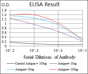
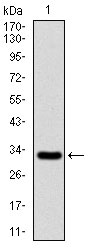
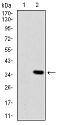
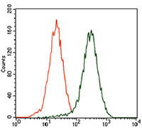
Application
| WB, FC, E |
|---|---|
| Primary Accession | Q6UVK1 |
| Reactivity | Human |
| Host | Mouse |
| Clonality | Monoclonal |
| Clone Names | 7G4E5 |
| Isotype | IgG1 |
| Calculated MW | 250537 Da |
| Description | A human melanoma-associated chondroitin sulfate proteoglycan plays a role in stabilizing cell-substratum interactions during early events of melanoma cell spreading on endothelial basement membranes. CSPG4 represents an integral membrane chondroitin sulfate proteoglycan expressed by human malignant melanoma cells. |
| Immunogen | Purified recombinant fragment of human CSPG4 (AA: 2247-2308) expressed in E. Coli. |
| Formulation | Purified antibody in PBS with 0.05% sodium azide |
| Gene ID | 1464 |
|---|---|
| Other Names | Chondroitin sulfate proteoglycan 4, Chondroitin sulfate proteoglycan NG2, Melanoma chondroitin sulfate proteoglycan, Melanoma-associated chondroitin sulfate proteoglycan, CSPG4, MCSP |
| Dilution | WB~~1/500 - 1/2000 FC~~1/200 - 1/400 E~~1/10000 |
| Storage | Maintain refrigerated at 2-8°C for up to 6 months. For long term storage store at -20°C in small aliquots to prevent freeze-thaw cycles. |
| Precautions | CSPG4 Antibody is for research use only and not for use in diagnostic or therapeutic procedures. |
| Name | CSPG4 |
|---|---|
| Synonyms | MCSP |
| Function | Proteoglycan playing a role in cell proliferation and migration which stimulates endothelial cells motility during microvascular morphogenesis. May also inhibit neurite outgrowth and growth cone collapse during axon regeneration. Cell surface receptor for collagen alpha 2(VI) which may confer cells ability to migrate on that substrate. Binds through its extracellular N-terminus growth factors, extracellular matrix proteases modulating their activity. May regulate MPP16-dependent degradation and invasion of type I collagen participating in melanoma cells invasion properties. May modulate the plasminogen system by enhancing plasminogen activation and inhibiting angiostatin. Also functions as a signal transducing protein by binding through its cytoplasmic C-terminus scaffolding and signaling proteins. May promote retraction fiber formation and cell polarization through Rho GTPase activation. May stimulate alpha-4, beta-1 integrin-mediated adhesion and spreading by recruiting and activating a signaling cascade through CDC42, ACK1 and BCAR1. May activate FAK and ERK1/ERK2 signaling cascades. |
| Cellular Location | Cell membrane {ECO:0000250|UniProtKB:Q00657}; Single-pass type I membrane protein {ECO:0000250|UniProtKB:Q00657}; Extracellular side {ECO:0000250|UniProtKB:Q00657}. Apical cell membrane {ECO:0000250|UniProtKB:Q00657}; Single-pass type I membrane protein {ECO:0000250|UniProtKB:Q00657}; Extracellular side {ECO:0000250|UniProtKB:Q00657}. Cell projection, lamellipodium membrane {ECO:0000250|UniProtKB:Q00657}; Single-pass type I membrane protein {ECO:0000250|UniProtKB:Q00657}; Extracellular side {ECO:0000250|UniProtKB:Q00657}. Cell surface {ECO:0000250|UniProtKB:Q00657}. Note=Localized at the apical plasma membrane it relocalizes to the lamellipodia of astrocytoma upon phosphorylation by PRKCA. Localizes to the retraction fibers. Localizes to the plasma membrane of oligodendrocytes (By similarity) {ECO:0000250|UniProtKB:Q00657, ECO:0000250|UniProtKB:Q8VHY0} |
| Tissue Location | Detected in fibroblasts (at protein level) (PubMed:36213313). Detected in placenta (at protein level) (PubMed:32337544). Detected in malignant melanoma cells |
For Research Use Only. Not For Use In Diagnostic Procedures.
Provided below are standard protocols that you may find useful for product applications.
BACKGROUND
This gene belongs to the RING finger family, members of which encode proteins characterized by a RING domain, a zinc-binding motif related to the zinc finger domain. The gene product can bind DNA and can act as a transcriptional repressor. It is associated with the multimeric polycomb group protein complex. The gene product interacts with the polycomb group proteins BMI1, EDR1, and CBX4, and colocalizes with these proteins in large nuclear domains. It interacts with the CBX4 protein via its glycine-rich C-terminal domain. The gene maps to the HLA class II region, where it is contiguous with the RING finger genes FABGL and HKE4.
REFERENCES
1.Cancer Res. 2011 Dec 15;71(24):7410-22. 2.Pigment Cell Melanoma Res. 2011 Dec;24(6):1148-57.
终于等到您。ABCEPTA(百远生物)抗体产品。
点击下方“我要评价 ”按钮提交您的反馈信息,您的反馈和评价是我们最宝贵的财富之一,
我们将在1-3个工作日内处理您的反馈信息。
如有疑问,联系:0512-88856768 tech-china@abcepta.com.


