


 癌症的基本特征包括细胞增殖、血管生成、迁移、凋亡逃避机制和细胞永生等。找到癌症发生过程中这些通路的关键标记物和对应的抗体用于检测至关重要。
 为您推荐一个泛素化位点预测神器——泛素化分析工具,可以为您的蛋白的泛素化位点作出预测和评分。
 细胞自噬受体图形绘图工具为你的蛋白的细胞受体结合位点作出预测和评分,识别结合到自噬通路中的蛋白是非常重要的,便于让我们理解自噬在正常生理、病理过程中的作用,如发育、细胞分化、神经退化性疾病、压力条件下、感染和癌症。
TFRC Antibody
Purified Mouse Monoclonal Antibody
- 产品详情
- 实验流程
- 背景知识
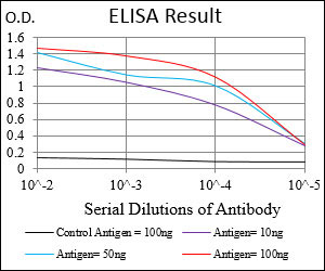
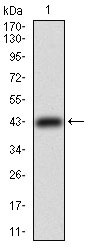
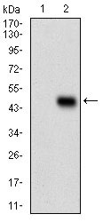
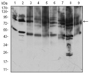
Application
| WB, E |
|---|---|
| Primary Accession | P02786 |
| Reactivity | Human, Mouse, Rat, Monkey |
| Host | Mouse |
| Clonality | Monoclonal |
| Clone Names | 1A1B2 |
| Isotype | IgG1 |
| Calculated MW | 84871 Da |
| Description | Transferrin receptor is a carrier protein for transferrin. It is needed for the import of iron into the cell and is regulated in response to intracellular iron concentration. Low iron concentrations promote increased levels of transferrin receptor, to increase iron intake into the cell. Thus, transferrin receptor maintains cellular iron homeostasis. Expression of human TFR1, but not human TFR2, in hamster cell lines markedly enhanced the infection of viruses pseudotyped with the glycoprotein of Machupo, Guanarito, and Junin viruses, but not with those of Lassa or lymphocytic choriomeningitis viruses. An anti-TFR1 antibody efficiently inhibited the replication of Machupo, Guanarito, Junin, and Sabia viruses, but not that of Lassa virus.TFR1 is a cellular receptor for New World hemorrhagic fever arenaviruses. |
| Immunogen | Purified recombinant fragment of human TFRC (AA: 608-727) expressed in E. Coli. |
| Formulation | Purified antibody in PBS with 0.05% sodium azide |
| Other Names | Transferrin receptor protein 1, TR, TfR, TfR1, Trfr, T9, p90, CD71, Transferrin receptor protein 1, serum form, sTfR, TFRC |
|---|---|
| Dilution | WB~~1/500 - 1/2000 E~~1/10000 |
| Storage | Maintain refrigerated at 2-8°C for up to 6 months. For long term storage store at -20°C in small aliquots to prevent freeze-thaw cycles. |
| Precautions | TFRC Antibody is for research use only and not for use in diagnostic or therapeutic procedures. |
For Research Use Only. Not For Use In Diagnostic Procedures.
Provided below are standard protocols that you may find useful for product applications.
BACKGROUND
The protein encoded by this gene is similar in sequence to 3'/5' exonucleolytic subunits of the RNA exosome. The exosome is a large multimeric ribonucleotide complex responsible for degrading various RNA substrates. Several transcript variants, some protein-coding and some not, have been found for this gene. ;
REFERENCES
1. Folia Histochem Cytobiol. 2012 Jul 5;50(2):304-11. 2. J Biol Chem. 2011 Oct 14;286(41):35708-15.
终于等到您。ABCEPTA(百远生物)抗体产品。
点击下方“我要评价 ”按钮提交您的反馈信息,您的反馈和评价是我们最宝贵的财富之一,
我们将在1-3个工作日内处理您的反馈信息。
如有疑问,联系:0512-88856768 tech-china@abcepta.com.


