


 癌症的基本特征包括细胞增殖、血管生成、迁移、凋亡逃避机制和细胞永生等。找到癌症发生过程中这些通路的关键标记物和对应的抗体用于检测至关重要。
 为您推荐一个泛素化位点预测神器——泛素化分析工具,可以为您的蛋白的泛素化位点作出预测和评分。
 细胞自噬受体图形绘图工具为你的蛋白的细胞受体结合位点作出预测和评分,识别结合到自噬通路中的蛋白是非常重要的,便于让我们理解自噬在正常生理、病理过程中的作用,如发育、细胞分化、神经退化性疾病、压力条件下、感染和癌症。
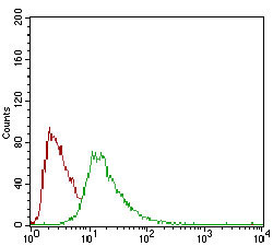
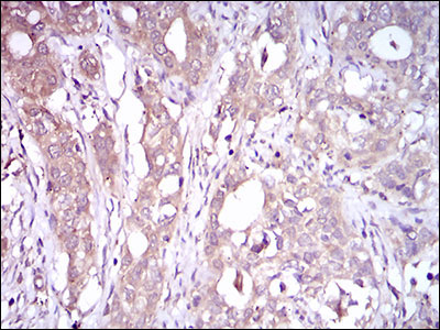
CD93 Antibody
Purified Mouse Monoclonal Antibody
- 产品详情
- 实验流程
- 背景知识
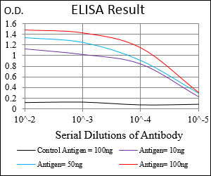
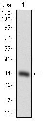
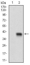
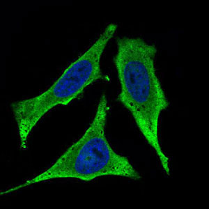
Application
| WB, IHC, FC, ICC, E |
|---|---|
| Primary Accession | Q9NPY3 |
| Reactivity | Human |
| Host | Mouse |
| Clonality | Monoclonal |
| Clone Names | 1A10E10 |
| Isotype | IgG1 |
| Calculated MW | 68560 Da |
| Description | The protein encoded by this gene is a cell-surface glycoprotein and type I membrane protein that was originally identified as a myeloid cell-specific marker. The encoded protein was once thought to be a receptor for C1q, but now is thought to instead be involved in intercellular adhesion and in the clearance of apoptotic cells. The intracellular cytoplasmic tail of this protein has been found to interact with moesin, a protein known to play a role in linking transmembrane proteins to the cytoskeleton and in the remodelling of the cytoskeleton. |
| Immunogen | Purified recombinant fragment of human CD93 (AA: 474-535) expressed in E. Coli. |
| Formulation | Ascitic fluid containing 0.03% sodium azide. |
| Gene ID | 22918 |
|---|---|
| Other Names | Complement component C1q receptor, C1q/MBL/SPA receptor, C1qR, C1qR(p), C1qRp, CDw93, Complement component 1 q subcomponent receptor 1, Matrix-remodeling-associated protein 4, CD93, CD93, C1QR1, MXRA4 |
| Dilution | WB~~1/500 - 1/2000 IHC~~1/200 - 1/1000 FC~~1/200 - 1/400 ICC~~N/A E~~1/10000 |
| Storage | Maintain refrigerated at 2-8°C for up to 6 months. For long term storage store at -20°C in small aliquots to prevent freeze-thaw cycles. |
| Precautions | CD93 Antibody is for research use only and not for use in diagnostic or therapeutic procedures. |
| Name | CD93 |
|---|---|
| Synonyms | C1QR1, MXRA4 |
| Function | Cell surface receptor that plays a role in various physiological processes including inflammation, phagocytosis, and cell adhesion. Plays a role in phagocytosis and enhances the uptake of apoptotic cells and immune complexes by acting as a receptor for defense collagens including surfactant protein A/SFTPA1, C1q, and mannose-binding lectin (MBL2) (PubMed:7977768). Plays a role in the regulation of endothelial cell function and adhesion by activating angiogenesis (PubMed:24809468). Mechanistically, exerts its angiogenic function by associating with beta-dystroglycan, leading to SRC- dependent phosphorylation and subsequent recruitment of CBL. In turn, CBL provides a docking site for downstream signaling components, such as CRKL to enhance cell migration (PubMed:26848865). Participates in angiogenesis also by acting as a receptor for the ECM pan-endothelial glycoprotein multimerin-2/MMRN2 and IGFBP7 ligands (PubMed:28671670, PubMed:36265539, PubMed:38218180). Both ligands play a non-redundant role in CD93-mediated endothelial cell function (PubMed:38218180). Acts as a key regulator of endothelial barrier function through modulating VEGFR2 function (By similarity). |
| Cellular Location | Cell membrane; Single-pass type I membrane protein |
| Tissue Location | Highly expressed in endothelial cells, platelets, cells of myeloid origin, such as monocytes and neutrophils. Not expressed in cells of lymphoid origin |
For Research Use Only. Not For Use In Diagnostic Procedures.
Provided below are standard protocols that you may find useful for product applications.
BACKGROUND
The enzyme encoded by this gene is an arylesterase that mainly hydrolyzes paroxon to produce p-nitrophenol. Paroxon is an organophosphorus anticholinesterase compound that is produced in vivo by oxidation of the insecticide parathion. Polymorphisms in this gene are a risk factor in coronary artery disease. The gene is found in a cluster of three related paraoxonase genes at 7q21.3. ; ; ;
REFERENCES
1. PLoS One. 2012;7(12):e51647. 2. J Clin Immunol. 2010 Sep;30(5):723-33.
终于等到您。ABCEPTA(百远生物)抗体产品。
点击下方“我要评价 ”按钮提交您的反馈信息,您的反馈和评价是我们最宝贵的财富之一,
我们将在1-3个工作日内处理您的反馈信息。
如有疑问,联系:0512-88856768 tech-china@abcepta.com.


