


 癌症的基本特征包括细胞增殖、血管生成、迁移、凋亡逃避机制和细胞永生等。找到癌症发生过程中这些通路的关键标记物和对应的抗体用于检测至关重要。
 为您推荐一个泛素化位点预测神器——泛素化分析工具,可以为您的蛋白的泛素化位点作出预测和评分。
 细胞自噬受体图形绘图工具为你的蛋白的细胞受体结合位点作出预测和评分,识别结合到自噬通路中的蛋白是非常重要的,便于让我们理解自噬在正常生理、病理过程中的作用,如发育、细胞分化、神经退化性疾病、压力条件下、感染和癌症。
DSG3 Antibody
Purified Mouse Monoclonal Antibody
- 产品详情
- 实验流程
- 背景知识
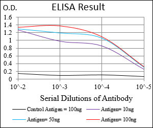
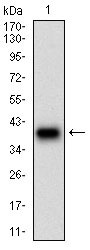
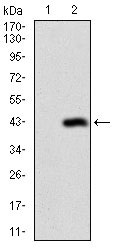
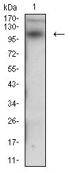
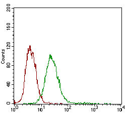
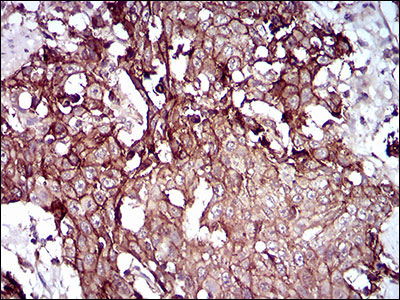
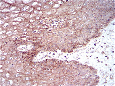
Application
| WB, IHC, FC, E |
|---|---|
| Primary Accession | P32926 |
| Reactivity | Human |
| Host | Mouse |
| Clonality | Monoclonal |
| Clone Names | 6G2C11 |
| Isotype | IgG1 |
| Calculated MW | 107533 Da |
| Description | Desmosomes are cell-cell junctions between epithelial, myocardial, and certain other cell types. Desmoglein 3 is a calcium-binding transmembrane glycoprotein component of desmosomes in vertebrate epithelial cells. Currently, three desmoglein subfamily members have been identified and all are members of the cadherin cell adhesion molecule superfamily. These desmoglein gene family members are located in a cluster on chromosome 18. This protein has been identified as the autoantigen of the autoimmune skin blistering disease pemphigus vulgaris. |
| Immunogen | Purified recombinant fragment of human DSG3 (AA: 55-159) expressed in E. Coli. |
| Formulation | Purified antibody in PBS with 0.05% sodium azide. |
| Gene ID | 1830 |
|---|---|
| Other Names | Desmoglein-3, 130 kDa pemphigus vulgaris antigen, PVA, Cadherin family member 6, DSG3, CDHF6 |
| Dilution | WB~~1/500 - 1/2000 IHC~~1/200 - 1/1000 FC~~1/200 - 1/400 E~~1/10000 |
| Storage | Maintain refrigerated at 2-8°C for up to 6 months. For long term storage store at -20°C in small aliquots to prevent freeze-thaw cycles. |
| Precautions | DSG3 Antibody is for research use only and not for use in diagnostic or therapeutic procedures. |
| Name | DSG3 (HGNC:3050) |
|---|---|
| Synonyms | CDHF6 |
| Function | A component of desmosome cell-cell junctions which are required for positive regulation of cellular adhesion (PubMed:31835537). Required for adherens and desmosome junction assembly in response to mechanical force in keratinocytes (PubMed:31835537). Required for desmosome-mediated cell-cell adhesion of cells surrounding the telogen hair club and the basal layer of the outer root sheath epithelium, consequently is essential for the anchoring of telogen hairs in the hair follicle (PubMed:9701552). Required for the maintenance of the epithelial barrier via promoting desmosome-mediated intercellular attachment of suprabasal epithelium to basal cells (By similarity). May play a role in the protein stability of the desmosome plaque components DSP, JUP, PKP1, PKP2 and PKP3 (PubMed:22294297). Required for YAP1 localization at the plasma membrane in keratinocytes in response to mechanical strain, via the formation of an interaction complex composed of DSG3, PKP1 and YWHAG (PubMed:31835537). May also be involved in the positive regulation of YAP1 target gene transcription and as a result cell proliferation (PubMed:31835537). Positively regulates cellular contractility and cell junction formation via organization of cortical F-actin bundles and anchoring of actin to tight junctions, in conjunction with RAC1 (PubMed:22796473). The cytoplasmic pool of DSG3 is required for the localization of CDH1 and CTNNB1 at developing adherens junctions, potentially via modulation of SRC activity (PubMed:22294297). Inhibits keratinocyte migration via suppression of p38MAPK signaling, may therefore play a role in moderating wound healing (PubMed:26763450). |
| Cellular Location | Cell membrane; Single-pass type I membrane protein. Cell junction, desmosome {ECO:0000250|UniProtKB:O35902}. Cytoplasm. Cell junction, tight junction. Cell junction |
| Tissue Location | Expressed throughout the basal and spinous layer of the epidermis with weak expression in the granular layer (at protein level) (PubMed:19717567). Expressed in skin and mucosa (at protein level) (PubMed:22294297, PubMed:30528827). Expressed in the basal layer of the outer root sheath of the telogen hair club, specifically at the cell membrane between the apex of the cells and the surrounding hair club (at protein level) (PubMed:9701552). Expression is less abundant between the lateral margins of the outer root sheath basal cells (at protein level) (PubMed:9701552). Also expressed in the tongue, tonsil and esophagus (PubMed:16740002). |
For Research Use Only. Not For Use In Diagnostic Procedures.
Provided below are standard protocols that you may find useful for product applications.
BACKGROUND
The protein encoded by this gene is found in the nucleus and is a cofactor of DNA polymerase delta. The encoded protein acts as a homotrimer and helps increase the processivity of leading strand synthesis during DNA replication. In response to DNA damage, this protein is ubiquitinated and is involved in the RAD6-dependent DNA repair pathway. Two transcript variants encoding the same protein have been found for this gene. Pseudogenes of this gene have been described on chromosome 4 and on the X chromosome. ; ;
REFERENCES
1. J Dermatol Sci. 2012 Feb;65(2):102-9. 2. Am J Pathol. 2009 May;174(5):1629-37.
终于等到您。ABCEPTA(百远生物)抗体产品。
点击下方“我要评价 ”按钮提交您的反馈信息,您的反馈和评价是我们最宝贵的财富之一,
我们将在1-3个工作日内处理您的反馈信息。
如有疑问,联系:0512-88856768 tech-china@abcepta.com.


