


 癌症的基本特征包括细胞增殖、血管生成、迁移、凋亡逃避机制和细胞永生等。找到癌症发生过程中这些通路的关键标记物和对应的抗体用于检测至关重要。
 为您推荐一个泛素化位点预测神器——泛素化分析工具,可以为您的蛋白的泛素化位点作出预测和评分。
 细胞自噬受体图形绘图工具为你的蛋白的细胞受体结合位点作出预测和评分,识别结合到自噬通路中的蛋白是非常重要的,便于让我们理解自噬在正常生理、病理过程中的作用,如发育、细胞分化、神经退化性疾病、压力条件下、感染和癌症。
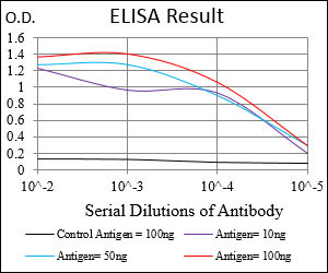
CAV2 Antibody
Purified Mouse Monoclonal Antibody
- 产品详情
- 实验流程
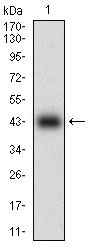
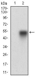
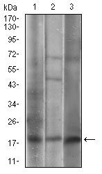
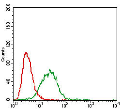
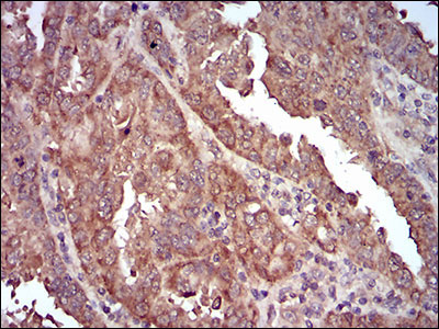
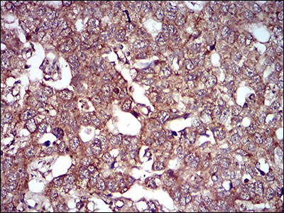
Application
| WB, IHC, FC, E |
|---|---|
| Primary Accession | P51636 |
| Reactivity | Human |
| Host | Mouse |
| Clonality | Monoclonal |
| Clone Names | 5E9E2 |
| Isotype | IgG1 |
| Calculated MW | 18291 Da |
| Description | The protein encoded by this gene is a major component of the inner surface of caveolae, small invaginations of the plasma membrane, and is involved in essential cellular functions, including signal transduction, lipid metabolism, cellular growth control and apoptosis. This protein may function as a tumor suppressor. This gene and related family member (CAV1) are located next to each other on chromosome 7, and express colocalizing proteins that form a stable hetero-oligomeric complex. Alternatively spliced transcript variants encoding different isoforms have been identified for this gene. Additional isoforms resulting from the use of alternate in-frame translation initiation codons have also been described, and shown to have preferential localization in the cell (PMID:11238462). |
| Immunogen | Purified recombinant fragment of human CAV2 (AA: 1-86) expressed in E. Coli. |
| Formulation | Purified antibody in PBS with 0.05% sodium azide. |
| Gene ID | 858 |
|---|---|
| Other Names | Caveolin-2, CAV2 |
| Dilution | WB~~1/500 - 1/2000 IHC~~1/200 - 1/1000 FC~~1/200 - 1/400 E~~1/10000 |
| Storage | Maintain refrigerated at 2-8°C for up to 6 months. For long term storage store at -20°C in small aliquots to prevent freeze-thaw cycles. |
| Precautions | CAV2 Antibody is for research use only and not for use in diagnostic or therapeutic procedures. |
| Name | CAV2 |
|---|---|
| Function | May act as a scaffolding protein within caveolar membranes. Interacts directly with G-protein alpha subunits and can functionally regulate their activity. Acts as an accessory protein in conjunction with CAV1 in targeting to lipid rafts and driving caveolae formation. The Ser-36 phosphorylated form has a role in modulating mitosis in endothelial cells. Positive regulator of cellular mitogenesis of the MAPK signaling pathway. Required for the insulin-stimulated nuclear translocation and activation of MAPK1 and STAT3, and the subsequent regulation of cell cycle progression (By similarity). |
| Cellular Location | Nucleus. Cytoplasm. Golgi apparatus membrane; Peripheral membrane protein. Cell membrane; Peripheral membrane protein. Membrane, caveola; Peripheral membrane protein. Note=Potential hairpin-like structure in the membrane. Membrane protein of caveolae Tyr-19-phosphorylated form is enriched at sites of cell-cell contact and is translocated to the nucleus in complex with MAPK1 in response to insulin (By similarity). Tyr-27-phosphorylated form is located both in the cytoplasm and plasma membrane. CAV1-mediated Ser-23-phosphorylated form locates to the plasma membrane. Ser-36-phosphorylated form resides in intracellular compartments. |
| Tissue Location | Expressed in endothelial cells, smooth muscle cells, skeletal myoblasts and fibroblasts |
Research Areas
For Research Use Only. Not For Use In Diagnostic Procedures.
Application Protocols
Provided below are standard protocols that you may find useful for product applications.
REFERENCES
1. Int J Oncol. 2011 May;38(5):1395-402.2. Breast Cancer Res Treat. 2008 Jul;110(2):245-56.
终于等到您。ABCEPTA(百远生物)抗体产品。
点击下方“我要评价 ”按钮提交您的反馈信息,您的反馈和评价是我们最宝贵的财富之一,
我们将在1-3个工作日内处理您的反馈信息。
如有疑问,联系:0512-88856768 tech-china@abcepta.com.
¥ 1,500.00
Cat# AO1935a


