


 癌症的基本特征包括细胞增殖、血管生成、迁移、凋亡逃避机制和细胞永生等。找到癌症发生过程中这些通路的关键标记物和对应的抗体用于检测至关重要。
 为您推荐一个泛素化位点预测神器——泛素化分析工具,可以为您的蛋白的泛素化位点作出预测和评分。
 细胞自噬受体图形绘图工具为你的蛋白的细胞受体结合位点作出预测和评分,识别结合到自噬通路中的蛋白是非常重要的,便于让我们理解自噬在正常生理、病理过程中的作用,如发育、细胞分化、神经退化性疾病、压力条件下、感染和癌症。
MLANA Antibody
Purified Mouse Monoclonal Antibody

- 产品详情
- 实验流程
- 背景知识
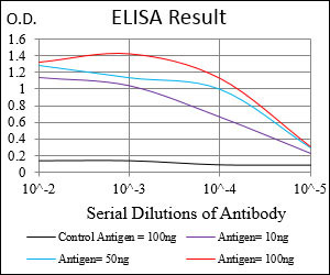
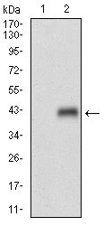
Application
| WB, E |
|---|---|
| Primary Accession | Q16655 |
| Reactivity | Human |
| Host | Mouse |
| Clonality | Monoclonal |
| Clone Names | 2F3B7 |
| Isotype | IgG1 |
| Calculated MW | 13157 Da |
| Description | MLANA (melan-A) is a protein-coding gene. Diseases associated with MLANA include meningeal melanocytoma, and juvenile xanthogranuloma. Involved in melanosome biogenesis by ensuring the stability of GPR143. Plays a vital role in the expression, stability, trafficking, and processing of melanocyte protein PMEL, which is critical to the formation of stage II melanosomes |
| Immunogen | Purified recombinant fragment of human MLANA (AA: 48-118) expressed in E. Coli. |
| Formulation | Purified antibody in PBS with 0.05% sodium azide. |
| Gene ID | 2315 |
|---|---|
| Other Names | Melanoma antigen recognized by T-cells 1, MART-1, Antigen LB39-AA, Antigen SK29-AA, Protein Melan-A, MLANA, MART1 |
| Dilution | WB~~1/500 - 1/2000 E~~1/10000 |
| Storage | Maintain refrigerated at 2-8°C for up to 6 months. For long term storage store at -20°C in small aliquots to prevent freeze-thaw cycles. |
| Precautions | MLANA Antibody is for research use only and not for use in diagnostic or therapeutic procedures. |
| Name | MLANA |
|---|---|
| Synonyms | MART1 |
| Function | Involved in melanosome biogenesis by ensuring the stability of GPR143. Plays a vital role in the expression, stability, trafficking, and processing of melanocyte protein PMEL, which is critical to the formation of stage II melanosomes. |
| Cellular Location | Endoplasmic reticulum membrane; Single-pass type III membrane protein. Golgi apparatus. Golgi apparatus, trans-Golgi network membrane. Melanosome. Note=Also found in small vesicles and tubules dispersed over the entire cytoplasm. A small fraction of the protein is inserted into the membrane in an inverted orientation Inversion of membrane topology results in the relocalization of the protein from a predominant Golgi/post-Golgi area to the endoplasmic reticulum. Melanoma cells expressing the protein with an inverted membrane topology are more effectively recognized by specific cytolytic T-lymphocytes than those expressing the protein in its native membrane orientation |
| Tissue Location | Expression is restricted to melanoma and melanocyte cell lines and retina |
For Research Use Only. Not For Use In Diagnostic Procedures.
Provided below are standard protocols that you may find useful for product applications.
BACKGROUND
The immunoglobulin epsilon receptor (IgE receptor) is the initiator of the allergic response. When two or more high-affinity IgE receptors are brought together by allergen-bound IgE molecules, mediators such as histamine that are responsible for allergy symptoms are released. This receptor is comprised of an alpha subunit, a beta subunit, and two gamma subunits. The protein encoded by this gene represents the alpha subunit. ; ; ;
REFERENCES
1. Mol Med Rep. 2011 Sep-Oct;4(5):799-803.2. J Cutan Pathol. 2011 Dec;38(12):954-60.
终于等到您。ABCEPTA(百远生物)抗体产品。
点击下方“我要评价 ”按钮提交您的反馈信息,您的反馈和评价是我们最宝贵的财富之一,
我们将在1-3个工作日内处理您的反馈信息。
如有疑问,联系:0512-88856768 tech-china@abcepta.com.


