


 癌症的基本特征包括细胞增殖、血管生成、迁移、凋亡逃避机制和细胞永生等。找到癌症发生过程中这些通路的关键标记物和对应的抗体用于检测至关重要。
 为您推荐一个泛素化位点预测神器——泛素化分析工具,可以为您的蛋白的泛素化位点作出预测和评分。
 细胞自噬受体图形绘图工具为你的蛋白的细胞受体结合位点作出预测和评分,识别结合到自噬通路中的蛋白是非常重要的,便于让我们理解自噬在正常生理、病理过程中的作用,如发育、细胞分化、神经退化性疾病、压力条件下、感染和癌症。
Neuropilin-1 Antibody
Purified Mouse Monoclonal Antibody
- 产品详情
- 实验流程
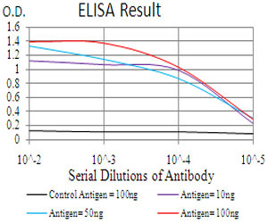
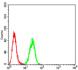
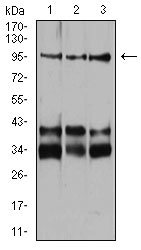
Application
| WB, FC, E |
|---|---|
| Primary Accession | O14786 |
| Reactivity | Human |
| Host | Mouse |
| Clonality | Monoclonal |
| Clone Names | 5B1E11 |
| Isotype | IgG1 |
| Calculated MW | 103134 Da |
| Description | This gene encodes one of two neuropilins, which contain specific protein domains which allow them to participate in several different types of signaling pathways that control cell migration. Neuropilins contain a large N-terminal extracellular domain, made up of complement-binding, coagulation factor V/VIII, and meprin domains. These proteins also contains a short membrane-spanning domain and a small cytoplasmic domain. Neuropilins bind many ligands and various types of co-receptors; they affect cell survival, migration, and attraction. Some of the ligands and co-receptors bound by neuropilins are vascular endothelial growth factor (VEGF) and semaphorin family members. Several alternatively spliced transcript variants that encode different protein isoforms have been described for this gene. |
| Immunogen | Synthesized peptide of human Neuropilin-1 (AA: 45-59). |
| Formulation | Purified antibody in PBS with 0.05% sodium azide |
| Gene ID | 8829 |
|---|---|
| Other Names | Neuropilin-1, Vascular endothelial cell growth factor 165 receptor, CD304, NRP1, NRP, VEGF165R |
| Dilution | WB~~1/500 - 1/2000 FC~~1/200 - 1/400 E~~1/10000 |
| Storage | Maintain refrigerated at 2-8°C for up to 6 months. For long term storage store at -20°C in small aliquots to prevent freeze-thaw cycles. |
| Precautions | Neuropilin-1 Antibody is for research use only and not for use in diagnostic or therapeutic procedures. |
| Name | NRP1 (HGNC:8004) |
|---|---|
| Synonyms | NRP, VEGF165R |
| Function | Cell-surface receptor involved in the development of the cardiovascular system, in angiogenesis, in the formation of certain neuronal circuits and in organogenesis outside the nervous system. Mediates the chemorepulsant activity of semaphorins (PubMed:10688880, PubMed:9288753, PubMed:9529250). Recognizes a C-end rule (CendR) motif R/KXXR/K on its ligands which causes cellular internalization and vascular leakage (PubMed:19805273). It binds to semaphorin 3A, the PLGF-2 isoform of PGF, the VEGF165 isoform of VEGFA and VEGFB (PubMed:10688880, PubMed:19805273, PubMed:9288753, PubMed:9529250). Coexpression with KDR results in increased VEGF165 binding to KDR as well as increased chemotaxis. Regulates VEGF-induced angiogenesis. Binding to VEGFA initiates a signaling pathway needed for motor neuron axon guidance and cell body migration, including for the caudal migration of facial motor neurons from rhombomere 4 to rhombomere 6 during embryonic development (By similarity). Regulates mitochondrial iron transport via interaction with ABCB8/MITOSUR (PubMed:30623799). |
| Cellular Location | [Isoform 2]: Secreted |
| Tissue Location | [Isoform 1]: The expression of isoforms 1 and 2 does not seem to overlap. Expressed in olfactory epithelium (at protein level) (PubMed:33082293). Expressed in fibroblasts (at protein level) (PubMed:36213313). Expressed by the blood vessels of different tissues In the developing embryo it is found predominantly in the nervous system. In adult tissues, it is highly expressed in heart and placenta; moderately in lung, liver, skeletal muscle, kidney and pancreas; and low in adult brain (PubMed:10688880, PubMed:9529250). Expressed in the central nervous system, including olfactory related regions such as the olfactory tubercles and paraolfactory gyri (PubMed:33082293) |
Research Areas
For Research Use Only. Not For Use In Diagnostic Procedures.
Application Protocols
Provided below are standard protocols that you may find useful for product applications.
REFERENCES
1.Leuk Res. 2012 Feb;36(2):169-73. 2.Blood. 2011 Jan 20;117(3):920-7.
终于等到您。ABCEPTA(百远生物)抗体产品。
点击下方“我要评价 ”按钮提交您的反馈信息,您的反馈和评价是我们最宝贵的财富之一,
我们将在1-3个工作日内处理您的反馈信息。
如有疑问,联系:0512-88856768 tech-china@abcepta.com.


