


 癌症的基本特征包括细胞增殖、血管生成、迁移、凋亡逃避机制和细胞永生等。找到癌症发生过程中这些通路的关键标记物和对应的抗体用于检测至关重要。
 为您推荐一个泛素化位点预测神器——泛素化分析工具,可以为您的蛋白的泛素化位点作出预测和评分。
 细胞自噬受体图形绘图工具为你的蛋白的细胞受体结合位点作出预测和评分,识别结合到自噬通路中的蛋白是非常重要的,便于让我们理解自噬在正常生理、病理过程中的作用,如发育、细胞分化、神经退化性疾病、压力条件下、感染和癌症。
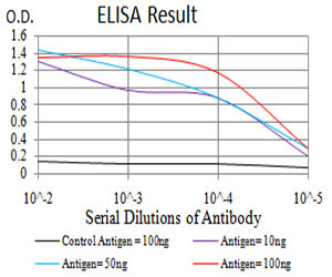
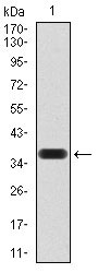
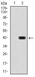
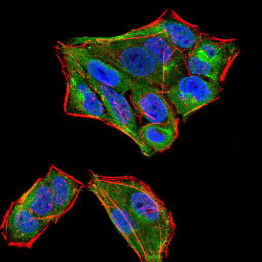
SIRT4 Antibody
Purified Mouse Monoclonal Antibody
- 产品详情
- 实验流程
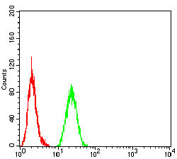
Application
| WB, FC, ICC, E |
|---|---|
| Primary Accession | Q9Y6E7 |
| Reactivity | Human |
| Host | Mouse |
| Clonality | Monoclonal |
| Clone Names | 6G6D2 |
| Isotype | IgG1 |
| Calculated MW | 35188 Da |
| Description | This gene encodes a member of the sirtuin family of proteins, homologs to the yeast Sir2 protein. Members of the sirtuin family are characterized by a sirtuin core domain and grouped into four classes. The functions of human sirtuins have not yet been determined; however, yeast sirtuin proteins are known to regulate epigenetic gene silencing and suppress recombination of rDNA. Studies suggest that the human sirtuins may function as intracellular regulatory proteins with mono-ADP-ribosyltransferase activity. The protein encoded by this gene is included in class IV of the sirtuin family. |
| Immunogen | Purified recombinant fragment of human SIRT4 (AA: 215-314) expressed in E. Coli. |
| Formulation | Purified antibody in PBS with 0.05% sodium azide |
| Gene ID | 23409 |
|---|---|
| Other Names | NAD-dependent protein deacetylase sirtuin-4 {ECO:0000255|HAMAP-Rule:MF_03161}, 3.5.1.- {ECO:0000255|HAMAP-Rule:MF_03161}, NAD-dependent ADP-ribosyltransferase sirtuin-4 {ECO:0000255|HAMAP-Rule:MF_03161}, 2.4.2.- {ECO:0000255|HAMAP-Rule:MF_03161}, Regulatory protein SIR2 homolog 4 {ECO:0000255|HAMAP-Rule:MF_03161}, SIR2-like protein 4 {ECO:0000255|HAMAP-Rule:MF_03161}, SIRT4 {ECO:0000255|HAMAP-Rule:MF_03161}, SIR2L4 |
| Dilution | WB~~1/500 - 1/2000 FC~~1/200 - 1/400 ICC~~N/A E~~1/10000 |
| Storage | Maintain refrigerated at 2-8°C for up to 6 months. For long term storage store at -20°C in small aliquots to prevent freeze-thaw cycles. |
| Precautions | SIRT4 Antibody is for research use only and not for use in diagnostic or therapeutic procedures. |
| Name | SIRT4 {ECO:0000255|HAMAP-Rule:MF_03161, ECO:0000312|HGNC:HGNC:14932} |
|---|---|
| Function | Acts as a NAD-dependent protein lipoamidase, biotinylase, deacetylase and ADP-ribosyl transferase (PubMed:16959573, PubMed:17715127, PubMed:24052263, PubMed:25525879). Catalyzes more efficiently removal of lipoyl- and biotinyl- than acetyl-lysine modifications (PubMed:24052263, PubMed:25525879). Inhibits the pyruvate dehydrogenase complex (PDH) activity via the enzymatic hydrolysis of the lipoamide cofactor from the E2 component, DLAT, in a phosphorylation-independent manner (PubMed:25525879). Catalyzes the transfer of ADP-ribosyl groups onto target proteins, including mitochondrial GLUD1, inhibiting GLUD1 enzyme activity (PubMed:16959573, PubMed:17715127). Acts as a negative regulator of mitochondrial glutamine metabolism by mediating mono ADP-ribosylation of GLUD1: expressed in response to DNA damage and negatively regulates anaplerosis by inhibiting GLUD1, leading to block metabolism of glutamine into tricarboxylic acid cycle and promoting cell cycle arrest (PubMed:16959573, PubMed:17715127). In response to mTORC1 signal, SIRT4 expression is repressed, promoting anaplerosis and cell proliferation (PubMed:23663782). Acts as a tumor suppressor (PubMed:23562301, PubMed:23663782). Also acts as a NAD-dependent protein deacetylase: mediates deacetylation of 'Lys-471' of MLYCD, inhibiting its activity, thereby acting as a regulator of lipid homeostasis (By similarity). Does not seem to deacetylate PC (PubMed:23438705). Controls fatty acid oxidation by inhibiting PPARA transcriptional activation (PubMed:24043310). Impairs SIRT1-PPARA interaction probably through the regulation of NAD(+) levels (PubMed:24043310). Down-regulates insulin secretion (PubMed:17715127). |
| Cellular Location | Mitochondrion matrix {ECO:0000255|HAMAP- Rule:MF_03161, ECO:0000269|PubMed:16079181, ECO:0000269|PubMed:16959573, ECO:0000269|PubMed:17715127} |
| Tissue Location | Detected in vascular smooth muscle and striated muscle. Detected in insulin-producing beta-cells in pancreas islets of Langerhans (at protein level). Widely expressed. Weakly expressed in leukocytes and fetal thymus. |
Research Areas
For Research Use Only. Not For Use In Diagnostic Procedures.
Application Protocols
Provided below are standard protocols that you may find useful for product applications.
REFERENCES
1.Eur J Histochem. 2011 Mar 21;55(1):e10.2.J Biol Chem. 2014 Feb 14;289(7):4135-44.
终于等到您。ABCEPTA(百远生物)抗体产品。
点击下方“我要评价 ”按钮提交您的反馈信息,您的反馈和评价是我们最宝贵的财富之一,
我们将在1-3个工作日内处理您的反馈信息。
如有疑问,联系:0512-88856768 tech-china@abcepta.com.


