


 癌症的基本特征包括细胞增殖、血管生成、迁移、凋亡逃避机制和细胞永生等。找到癌症发生过程中这些通路的关键标记物和对应的抗体用于检测至关重要。
 为您推荐一个泛素化位点预测神器——泛素化分析工具,可以为您的蛋白的泛素化位点作出预测和评分。
 细胞自噬受体图形绘图工具为你的蛋白的细胞受体结合位点作出预测和评分,识别结合到自噬通路中的蛋白是非常重要的,便于让我们理解自噬在正常生理、病理过程中的作用,如发育、细胞分化、神经退化性疾病、压力条件下、感染和癌症。
CDH5 Antibody
Purified Mouse Monoclonal Antibody
- 产品详情
- 实验流程
Application
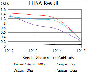
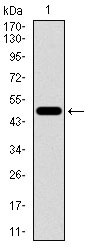
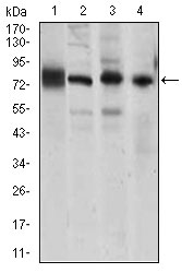
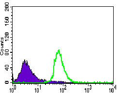
| WB, FC, E |
|---|---|
| Primary Accession | P33151 |
| Reactivity | Human, Mouse |
| Host | Mouse |
| Clonality | Monoclonal |
| Clone Names | 3D5C7 |
| Isotype | IgG1 |
| Calculated MW | 87528 Da |
| Description | This gene is a classical cadherin from the cadherin superfamily and is located in a six-cadherin cluster in a region on the long arm of chromosome 16 that is involved in loss of heterozygosity events in breast and prostate cancer. The encoded protein is a calcium-dependent cell-cell adhesion glycoprotein comprised of five extracellular cadherin repeats, a transmembrane region and a highly conserved cytoplasmic tail. Functioning as a classic cadherin by imparting to cells the ability to adhere in a homophilic manner, the protein may play an important role in endothelial cell biology through control of the cohesion and organization of the intercellular junctions. An alternative splice variant has been described but its full length sequence has not been determined. |
| Immunogen | Purified recombinant fragment of human CDH5 (AA: 29-223 ) expressed in E. Coli. |
| Formulation | Ascitic fluid containing 0.03% sodium azide. |
| Other Names | Cadherin-5, 7B4 antigen, Vascular endothelial cadherin, VE-cadherin, CD144, CDH5 |
|---|---|
| Dilution | WB~~1/500 - 1/2000 FC~~1/200 - 1/400 E~~1/10000 |
| Storage | Maintain refrigerated at 2-8°C for up to 6 months. For long term storage store at -20°C in small aliquots to prevent freeze-thaw cycles. |
| Precautions | CDH5 Antibody is for research use only and not for use in diagnostic or therapeutic procedures. |
Research Areas
For Research Use Only. Not For Use In Diagnostic Procedures.
Application Protocols
Provided below are standard protocols that you may find useful for product applications.
REFERENCES
1.Blood. 2011 Feb 24;117(8):2515-26. 2.J Virol. 2010 Nov;84(21):11227-34.
终于等到您。ABCEPTA(百远生物)抗体产品。
点击下方“我要评价 ”按钮提交您的反馈信息,您的反馈和评价是我们最宝贵的财富之一,
我们将在1-3个工作日内处理您的反馈信息。
如有疑问,联系:0512-88856768 tech-china@abcepta.com.
¥ 1,520.00
Cat# AO2248a


