


 癌症的基本特征包括细胞增殖、血管生成、迁移、凋亡逃避机制和细胞永生等。找到癌症发生过程中这些通路的关键标记物和对应的抗体用于检测至关重要。
 为您推荐一个泛素化位点预测神器——泛素化分析工具,可以为您的蛋白的泛素化位点作出预测和评分。
 细胞自噬受体图形绘图工具为你的蛋白的细胞受体结合位点作出预测和评分,识别结合到自噬通路中的蛋白是非常重要的,便于让我们理解自噬在正常生理、病理过程中的作用,如发育、细胞分化、神经退化性疾病、压力条件下、感染和癌症。
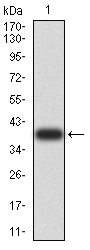
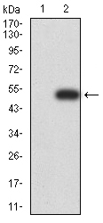
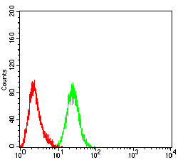
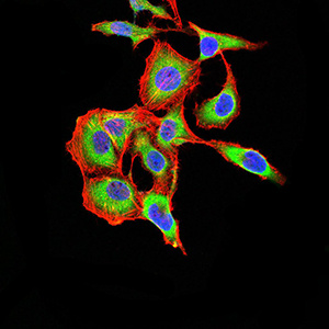
Mouse Monoclonal Antibody to ARF1
Purified Mouse Monoclonal Antibody


- 产品详情
- 实验流程
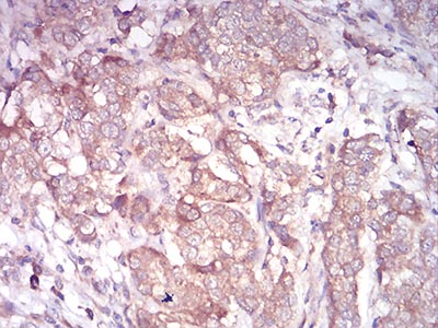
Application
| WB, IHC, FC, ICC, E |
|---|---|
| Primary Accession | P84077 |
| Reactivity | Human |
| Host | Mouse |
| Clonality | Monoclonal |
| Clone Names | 8D8E9 |
| Isotype | Mouse IgG2a |
| Calculated MW | 20697 Da |
| Description | ADP-ribosylation factor 1 (ARF1) is a member of the human ARF gene family. The family members encode small guanine nucleotide-binding proteins that stimulate the ADP-ribosyltransferase activity of cholera toxin and play a role in vesicular trafficking as activators of phospholipase D. The gene products, including 6 ARF proteins and 11 ARF-like proteins, constitute a family of the RAS superfamily. The ARF proteins are categorized as class I (ARF1, ARF2 and ARF3), class II (ARF4 and ARF5) and class III (ARF6), and members of each class share a common gene organization. The ARF1 protein is localized to the Golgi apparatus and has a central role in intra-Golgi transport. Multiple alternatively spliced transcript variants encoding the same protein have been found for this gene.; |
| Immunogen | Purified recombinant fragment of human ARF1 (AA: 76-182) expressed in E. Coli. |
| Formulation | Purified antibody in PBS with 0.05% sodium azide |
| Application Note | ELISA: 1/10000; WB: 1/500 - 1/2000; IHC: 1/200 - 1/1000; ICC: 1/200 - 1/1000; FCM: 1/200 - 1/400 |
| Gene ID | 375 |
|---|---|
| Other Names | ADP-ribosylation factor 1, ARF1 |
| Dilution | WB~~1:1000 IHC~~1:100~500 FC~~1:10~50 ICC~~N/A E~~N/A |
| Storage | Maintain refrigerated at 2-8°C for up to 6 months. For long term storage store at -20°C in small aliquots to prevent freeze-thaw cycles. |
| Precautions | Mouse Monoclonal Antibody to ARF1 is for research use only and not for use in diagnostic or therapeutic procedures. |
| Name | ARF1 |
|---|---|
| Function | Small GTPase involved in protein trafficking between different compartments (PubMed:8253837). Modulates vesicle budding and uncoating within the Golgi complex (PubMed:8253837). In its GTP-bound form, triggers the recruitment of coatomer proteins to the Golgi membrane (PubMed:8253837). The hydrolysis of ARF1-bound GTP, which is mediated by ARFGAPs proteins, is required for dissociation of coat proteins from Golgi membranes and vesicles (PubMed:8253837). The GTP- bound form interacts with PICK1 to limit PICK1-mediated inhibition of Arp2/3 complex activity; the function is linked to AMPA receptor (AMPAR) trafficking, regulation of synaptic plasticity of excitatory synapses and spine shrinkage during long-term depression (LTD) (By similarity). Plays a key role in the regulation of intestinal stem cells and gut microbiota, and is essential for maintaining intestinal homeostasis (By similarity). Also plays a critical role in mast cell expansion but not in mast cell maturation by facilitating optimal mTORC1 activation (By similarity). |
| Cellular Location | Golgi apparatus membrane; Lipid-anchor; Cytoplasmic side. Synapse, synaptosome {ECO:0000250|UniProtKB:P84079}. Postsynaptic density {ECO:0000250|UniProtKB:P84079}. Note=In the GDP-bound form, associates transiently with the membranes via its myristoylated N-terminus where guanine nucleotide-exchange factor (GEF)-mediated nucleotide exchange occurs (By similarity). Following nucleotide exchange, the GTP-bound form undergoes a conformational change, leading to the exposure of a myristoylated N-terminal amphipathic helix that provides stable membrane anchorage (By similarity). {ECO:0000250|UniProtKB:P84080} |
Research Areas
For Research Use Only. Not For Use In Diagnostic Procedures.
Application Protocols
Provided below are standard protocols that you may find useful for product applications.
REFERENCES
1.Mol Biol Cell. 2014 Jan;25(1):17-29. ; 2.Cancer Sci. 2012 Jun;103(6):1136-44. ;
终于等到您。ABCEPTA(百远生物)抗体产品。
点击下方“我要评价 ”按钮提交您的反馈信息,您的反馈和评价是我们最宝贵的财富之一,
我们将在1-3个工作日内处理您的反馈信息。
如有疑问,联系:0512-88856768 tech-china@abcepta.com.
¥ 1,500.00
Cat# AO2352a


