


 癌症的基本特征包括细胞增殖、血管生成、迁移、凋亡逃避机制和细胞永生等。找到癌症发生过程中这些通路的关键标记物和对应的抗体用于检测至关重要。
 为您推荐一个泛素化位点预测神器——泛素化分析工具,可以为您的蛋白的泛素化位点作出预测和评分。
 细胞自噬受体图形绘图工具为你的蛋白的细胞受体结合位点作出预测和评分,识别结合到自噬通路中的蛋白是非常重要的,便于让我们理解自噬在正常生理、病理过程中的作用,如发育、细胞分化、神经退化性疾病、压力条件下、感染和癌症。
Mouse Monoclonal Antibody to TLR9
Purified Mouse Monoclonal Antibody
- 产品详情
- 实验流程
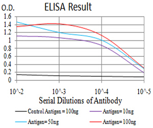
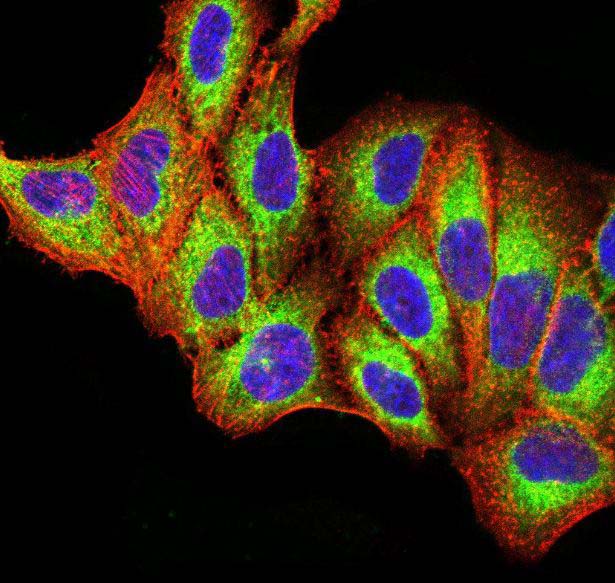
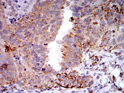
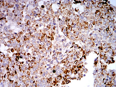
Application
| WB, IHC, FC, ICC, E |
|---|---|
| Primary Accession | Q9NR96 |
| Reactivity | Human |
| Host | Mouse |
| Clonality | Monoclonal |
| Clone Names | 1B12H2 |
| Isotype | Mouse IgG2a |
| Calculated MW | 115860 Da |
| Description | The protein encoded by this gene is a member of the Toll-like receptor (TLR) family which plays a fundamental role in pathogen recognition and activation of innate immunity. TLRs are highly conserved from Drosophila to humans and share structural and functional similarities. They recognize pathogen-associated molecular patterns (PAMPs) that are expressed on infectious agents, and mediate the production of cytokines necessary for the development of effective immunity. The various TLRs exhibit different patterns of expression. This gene is preferentially expressed in immune cell rich tissues, such as spleen, lymph node, bone marrow and peripheral blood leukocytes. Studies in mice and human indicate that this receptor mediates cellular response to unmethylated CpG dinucleotides in bacterial DNA to mount an innate immune response.; |
| Immunogen | Purified recombinant fragment of human TLR9 (AA: 868-1016) expressed in E. Coli. |
| Formulation | Purified antibody in PBS with 0.05% sodium azide |
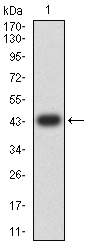
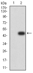
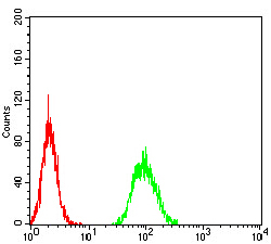
| Application Note | ELISA: 1/10000; WB: 1/500 - 1/2000; IHC: 1/200 - 1/1000; ICC: 1/200 - 1/1000; FCM: 1/200 - 1/400 |
| Other Names | CD289 |
|---|---|
| Dilution | WB~~1:1000 IHC~~1:100~500 FC~~1:10~50 ICC~~N/A E~~N/A |
| Storage | Maintain refrigerated at 2-8°C for up to 6 months. For long term storage store at -20°C in small aliquots to prevent freeze-thaw cycles. |
| Precautions | Mouse Monoclonal Antibody to TLR9 is for research use only and not for use in diagnostic or therapeutic procedures. |
Research Areas
For Research Use Only. Not For Use In Diagnostic Procedures.
Application Protocols
Provided below are standard protocols that you may find useful for product applications.
REFERENCES
1.J Virol. 2015 Nov;89(22):11396-405. ; 2.Immunogenetics. 2014 Dec;66(12):675-81.;
终于等到您。ABCEPTA(百远生物)抗体产品。
点击下方“我要评价 ”按钮提交您的反馈信息,您的反馈和评价是我们最宝贵的财富之一,
我们将在1-3个工作日内处理您的反馈信息。
如有疑问,联系:0512-88856768 tech-china@abcepta.com.
¥ 1,500.00
Cat# AO2370a


