


 癌症的基本特征包括细胞增殖、血管生成、迁移、凋亡逃避机制和细胞永生等。找到癌症发生过程中这些通路的关键标记物和对应的抗体用于检测至关重要。
 为您推荐一个泛素化位点预测神器——泛素化分析工具,可以为您的蛋白的泛素化位点作出预测和评分。
 细胞自噬受体图形绘图工具为你的蛋白的细胞受体结合位点作出预测和评分,识别结合到自噬通路中的蛋白是非常重要的,便于让我们理解自噬在正常生理、病理过程中的作用,如发育、细胞分化、神经退化性疾病、压力条件下、感染和癌症。
Mouse Monoclonal Antibody to MIB1
Purified Mouse Monoclonal Antibody
- 产品详情
- 实验流程
Application
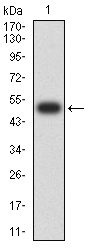
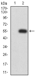
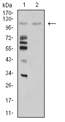
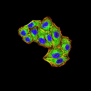
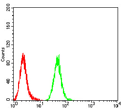
| WB, FC, ICC, E |
|---|---|
| Primary Accession | Q86YT6 |
| Reactivity | Human, Monkey |
| Host | Mouse |
| Clonality | Monoclonal |
| Clone Names | 6A9C9 |
| Isotype | Mouse IgG1 |
| Calculated MW | 110136 Da |
| Description | This gene encodes a protein containing multiple ankyrin repeats and RING finger domains that functions as an E3 ubiquitin ligase. The encoded protein positively regulates Notch signaling by ubiquitinating the Notch receptors, thereby facilitating their endocytosis. This protein may also promote the ubiquitination and degradation of death-associated protein kinase 1 (DAPK1). ; |
| Immunogen | Purified recombinant fragment of human MIB1 (AA: 6-221) expressed in E. Coli. |
| Formulation | Purified antibody in PBS with 0.05% sodium azide |
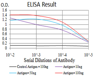
| Application Note | ELISA: 1/10000; WB: 1/500 - 1/2000; ICC: 1/200 - 1/1000; FCM: 1/200 - 1/400 |
| Gene ID | 57534 |
|---|---|
| Other Names | MIB; DIP1; ZZZ6; DIP-1; LVNC7; ZZANK2 |
| Dilution | WB~~1:1000 FC~~1:10~50 ICC~~N/A E~~N/A |
| Storage | Maintain refrigerated at 2-8°C for up to 6 months. For long term storage store at -20°C in small aliquots to prevent freeze-thaw cycles. |
| Precautions | Mouse Monoclonal Antibody to MIB1 is for research use only and not for use in diagnostic or therapeutic procedures. |
| Name | MIB1 |
|---|---|
| Synonyms | DIP1, KIAA1323, ZZANK2 |
| Function | E3 ubiquitin-protein ligase that mediates ubiquitination of Delta receptors, which act as ligands of Notch proteins. Positively regulates the Delta-mediated Notch signaling by ubiquitinating the intracellular domain of Delta, leading to endocytosis of Delta receptors. Probably mediates ubiquitination and subsequent proteasomal degradation of DAPK1, thereby antagonizing anti-apoptotic effects of DAPK1 to promote TNF-induced apoptosis (By similarity). Involved in ubiquitination of centriolar satellite CEP131, CEP290 and PCM1 proteins and hence inhibits primary cilium formation in proliferating cells. Mediates 'Lys-63'-linked polyubiquitination of TBK1, which probably participates in kinase activation. |
| Cellular Location | Cytoplasm. Cytoplasm, cytoskeleton, microtubule organizing center, centrosome, centriolar satellite. Cell membrane. Note=Localizes to the plasma membrane (By similarity) According to PubMed:15048887, it is mitochondrial, however such localization remains unclear. Displaced from centriolar satellites in response to cellular stress, such as ultraviolet light (UV) radiation or heat shock. |
| Tissue Location | Widely expressed at low level. Expressed at higher level in spinal cord, ovary, whole brain, and all specific brain regions examined. |
Research Areas
For Research Use Only. Not For Use In Diagnostic Procedures.
Application Protocols
Provided below are standard protocols that you may find useful for product applications.
REFERENCES
1.J Cell Sci. 2015 May 1;128(9):1674-82. ; 2.Cell Res. 2012 Mar;22(3):603-6. ;
终于等到您。ABCEPTA(百远生物)抗体产品。
点击下方“我要评价 ”按钮提交您的反馈信息,您的反馈和评价是我们最宝贵的财富之一,
我们将在1-3个工作日内处理您的反馈信息。
如有疑问,联系:0512-88856768 tech-china@abcepta.com.
¥ 1,500.00
Cat# AO2429a


