


 癌症的基本特征包括细胞增殖、血管生成、迁移、凋亡逃避机制和细胞永生等。找到癌症发生过程中这些通路的关键标记物和对应的抗体用于检测至关重要。
 为您推荐一个泛素化位点预测神器——泛素化分析工具,可以为您的蛋白的泛素化位点作出预测和评分。
 细胞自噬受体图形绘图工具为你的蛋白的细胞受体结合位点作出预测和评分,识别结合到自噬通路中的蛋白是非常重要的,便于让我们理解自噬在正常生理、病理过程中的作用,如发育、细胞分化、神经退化性疾病、压力条件下、感染和癌症。
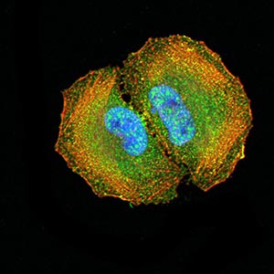
Mouse Monoclonal Antibody to DOC2
Purified Mouse Monoclonal Antibody
- 产品详情
- 实验流程
Application
| WB, FC, ICC, E |
|---|---|
| Primary Accession | P98082 |
| Reactivity | Human |
| Host | Mouse |
| Clonality | Monoclonal |
| Clone Names | 2H7C4 |
| Isotype | Mouse IgG1 |
| Calculated MW | 82448 Da |
| Description | This gene encodes a mitogen-responsive phosphoprotein. It is expressed in normal ovarian epithelial cells, but is down-regulated or absent from ovarian carcinoma cell lines, suggesting its role as a tumor suppressor. This protein binds to the SH3 domains of GRB2, an adaptor protein that couples tyrosine kinase receptors to SOS (a guanine nucleotide exchange factor for Ras), via its C-terminal proline-rich sequences, and may thus modulate growth factor/Ras pathways by competing with SOS for binding to GRB2. Alternatively spliced transcript variants encoding different isoforms have been found for this gene.; |
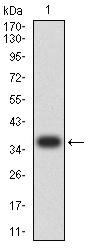
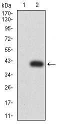
| Immunogen | Purified recombinant fragment of human DOC2 (AA: 652-749) expressed in E. Coli. |
| Formulation | Purified antibody in PBS with 0.05% sodium azide |
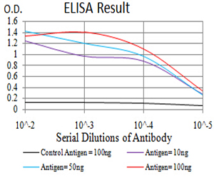
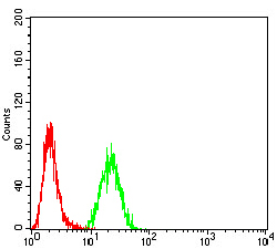
| Application Note | ELISA: 1/10000; WB: 1/500 - 1/2000; ICC: 1/200 - 1/1000; FCM: 1/200 - 1/400 |
| Gene ID | 1601 |
|---|---|
| Other Names | DAB2; DOC-2 |
| Dilution | WB~~1:1000 FC~~1:10~50 ICC~~N/A E~~N/A |
| Storage | Maintain refrigerated at 2-8°C for up to 6 months. For long term storage store at -20°C in small aliquots to prevent freeze-thaw cycles. |
| Precautions | Mouse Monoclonal Antibody to DOC2 is for research use only and not for use in diagnostic or therapeutic procedures. |
| Name | DAB2 |
|---|---|
| Synonyms | DOC2 |
| Function | Adapter protein that functions as a clathrin-associated sorting protein (CLASP) required for clathrin-mediated endocytosis of selected cargo proteins. Can bind and assemble clathrin, and binds simultaneously to phosphatidylinositol 4,5-bisphosphate (PtdIns(4,5)P2) and cargos containing non-phosphorylated NPXY internalization motifs, such as the LDL receptor, to recruit them to clathrin-coated pits. Can function in clathrin-mediated endocytosis independently of the AP-2 complex. Involved in endocytosis of integrin beta-1; this function seems to redundant with the AP-2 complex and seems to require DAB2 binding to endocytosis accessory EH domain-containing proteins such as EPS15, EPS15L1 and ITSN1. Involved in endocytosis of cystic fibrosis transmembrane conductance regulator/CFTR. Involved in endocytosis of megalin/LRP2 lipoprotein receptor during embryonal development. Required for recycling of the TGF-beta receptor. Involved in CFTR trafficking to the late endosome. Involved in several receptor-mediated signaling pathways. Involved in TGF-beta receptor signaling and facilitates phosphorylation of the signal transducer SMAD2. Mediates TFG-beta-stimulated JNK activation. May inhibit the canoniocal Wnt/beta-catenin signaling pathway by stabilizing the beta-catenin destruction complex through a competing association with axin preventing its dephosphorylation through protein phosphatase 1 (PP1). Sequesters LRP6 towards clathrin-mediated endocytosis, leading to inhibition of Wnt/beta-catenin signaling. May activate non-canonical Wnt signaling. In cell surface growth factor/Ras signaling pathways proposed to inhibit ERK activation by interrupting the binding of GRB2 to SOS1 and to inhibit SRC by preventing its activating phosphorylation at 'Tyr-419'. Proposed to be involved in modulation of androgen receptor (AR) signaling mediated by SRC activation; seems to compete with AR for interaction with SRC. Plays a role in the CSF-1 signal transduction pathway. Plays a role in cellular differentiation. Involved in cell positioning and formation of visceral endoderm (VE) during embryogenesis and proposed to be required in the VE to respond to Nodal signaling coming from the epiblast. Required for the epithelial to mesenchymal transition, a process necessary for proper embryonic development. May be involved in myeloid cell differentiation and can induce macrophage adhesion and spreading. May act as a tumor suppressor. |
| Cellular Location | Cytoplasm. Cytoplasmic vesicle, clathrin-coated vesicle membrane. Membrane, clathrin-coated pit. Note=Colocalizes with large insert-containing isoforms of MYO6 at clathrin-coated pits/vesicles. During mitosis is progressively displaced from the membrane and translocated to the cytoplasm |
| Tissue Location | Expressed in deep invaginations, inclusion cysts and the surface epithelial cells of the ovary. Also expressed in breast epithelial cells, spleen, thymus, prostate, testis, macrophages, fibroblasts, lung epithelial cells, placenta, brain stem, heart and small intestine. Expressed in kidney proximal tubular epithelial cells (at protein level). |
Research Areas
For Research Use Only. Not For Use In Diagnostic Procedures.
Application Protocols
Provided below are standard protocols that you may find useful for product applications.
REFERENCES
1.Mol Biol Cell. 2014 May;25(10):1620-8. ; 2.Exp Cell Res. 2012 Mar 10;318(5):550-7. ;
终于等到您。ABCEPTA(百远生物)抗体产品。
点击下方“我要评价 ”按钮提交您的反馈信息,您的反馈和评价是我们最宝贵的财富之一,
我们将在1-3个工作日内处理您的反馈信息。
如有疑问,联系:0512-88856768 tech-china@abcepta.com.


