


 癌症的基本特征包括细胞增殖、血管生成、迁移、凋亡逃避机制和细胞永生等。找到癌症发生过程中这些通路的关键标记物和对应的抗体用于检测至关重要。
 为您推荐一个泛素化位点预测神器——泛素化分析工具,可以为您的蛋白的泛素化位点作出预测和评分。
 细胞自噬受体图形绘图工具为你的蛋白的细胞受体结合位点作出预测和评分,识别结合到自噬通路中的蛋白是非常重要的,便于让我们理解自噬在正常生理、病理过程中的作用,如发育、细胞分化、神经退化性疾病、压力条件下、感染和癌症。
Mouse Monoclonal Antibody to ATG2A
Purified Mouse Monoclonal Antibody


- 产品详情
- 实验流程
Application
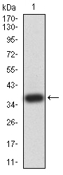
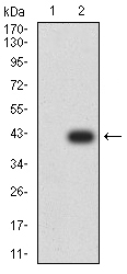
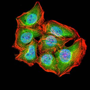
| WB, FC, ICC, E |
|---|---|
| Primary Accession | Q2TAZ0 |
| Reactivity | Human |
| Host | Mouse |
| Clonality | Monoclonal |
| Clone Names | 4E6D4 |
| Isotype | Mouse IgG1 |
| Calculated MW | 212860 Da |
| Description | ATG2A (Autophagy Related 2A) is a Protein Coding gene. An important paralog of this gene is ATG2B.; |
| Immunogen | Purified recombinant fragment of human ATG2A (AA: 325-429) expressed in E. Coli. |
| Formulation | Purified antibody in PBS with 0.05% sodium azide |
| Application Note | ELISA: 1/10000; WB: 1/500 - 1/2000; ICC: 1/50- 1/200; FCM: 1/200 - 1/400 |
| Gene ID | 23130 |
|---|---|
| Other Names | Autophagy-related protein 2 homolog A, ATG2A, KIAA0404 |
| Dilution | WB~~1:1000 FC~~1:10~50 ICC~~N/A E~~N/A |
| Storage | Maintain refrigerated at 2-8°C for up to 6 months. For long term storage store at -20°C in small aliquots to prevent freeze-thaw cycles. |
| Precautions | Mouse Monoclonal Antibody to ATG2A is for research use only and not for use in diagnostic or therapeutic procedures. |
| Name | ATG2A {ECO:0000303|PubMed:21887408, ECO:0000312|HGNC:HGNC:29028} |
|---|---|
| Function | Lipid transfer protein involved in autophagosome assembly (PubMed:28561066, PubMed:30952800, PubMed:31271352). Tethers the edge of the isolation membrane (IM) to the endoplasmic reticulum (ER) and mediates direct lipid transfer from ER to IM for IM expansion (PubMed:30952800, PubMed:31271352). Binds to the ER exit site (ERES), which is the membrane source for autophagosome formation, and extracts phospholipids from the membrane source and transfers them to ATG9 (ATG9A or ATG9B) to the IM for membrane expansion (PubMed:30952800, PubMed:31271352). Lipid transfer activity is enhanced by WIPI1 and WDR45/WIPI4, which promote ATG2A-association with phosphatidylinositol 3-monophosphate (PI3P)-containing membranes (PubMed:31271352). Also regulates lipid droplets morphology and distribution within the cell (PubMed:22219374, PubMed:28561066). |
| Cellular Location | Preautophagosomal structure membrane; Peripheral membrane protein {ECO:0000250|UniProtKB:Q96BY7}. Lipid droplet {ECO:0000250|UniProtKB:Q96BY7}. Endoplasmic reticulum membrane; Peripheral membrane protein {ECO:0000250|UniProtKB:P53855}. Note=Localizes to endoplasmic reticulum-autophagosome contact sites. |
Research Areas
For Research Use Only. Not For Use In Diagnostic Procedures.
Application Protocols
Provided below are standard protocols that you may find useful for product applications.
REFERENCES
1.Mol Biol Cell. 2012 Mar;23(5):896-909. ; 2.Acta Biochim Pol. 2011;58(3):365-74.;
终于等到您。ABCEPTA(百远生物)抗体产品。
点击下方“我要评价 ”按钮提交您的反馈信息,您的反馈和评价是我们最宝贵的财富之一,
我们将在1-3个工作日内处理您的反馈信息。
如有疑问,联系:0512-88856768 tech-china@abcepta.com.


