


 癌症的基本特征包括细胞增殖、血管生成、迁移、凋亡逃避机制和细胞永生等。找到癌症发生过程中这些通路的关键标记物和对应的抗体用于检测至关重要。
 为您推荐一个泛素化位点预测神器——泛素化分析工具,可以为您的蛋白的泛素化位点作出预测和评分。
 细胞自噬受体图形绘图工具为你的蛋白的细胞受体结合位点作出预测和评分,识别结合到自噬通路中的蛋白是非常重要的,便于让我们理解自噬在正常生理、病理过程中的作用,如发育、细胞分化、神经退化性疾病、压力条件下、感染和癌症。
BAG1
Purified Mouse Monoclonal Antibody
- 产品详情
- 实验流程
Application
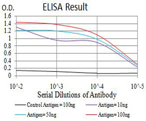
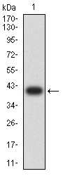
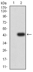
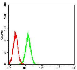
| WB, IHC, ICC, E |
|---|---|
| Primary Accession | Q99933 |
| Reactivity | Human |
| Host | Mouse |
| Clonality | Monoclonal |
| Clone Names | 2F7A11 |
| Isotype | Mouse IgG1 |
| Calculated MW | 38779 Da |
| Immunogen | Purified recombinant fragment of human BAG1 (AA: 219-346) expressed in E. Coli. |
| Formulation | Purified antibody in PBS with 0.05% sodium azide |
| Gene ID | 573 |
|---|---|
| Other Names | HAP; BAG-1; RAP46 |
| Dilution | WB~~ 1/500 - 1/2000 IHC~~1:100~500 ICC~~N/A E~~ 1/10000 |
| Storage | Maintain refrigerated at 2-8°C for up to 6 months. For long term storage store at -20°C in small aliquots to prevent freeze-thaw cycles. |
| Precautions | BAG1 is for research use only and not for use in diagnostic or therapeutic procedures. |
| Name | BAG1 |
|---|---|
| Synonyms | HAP |
| Function | Co-chaperone for HSP70 and HSC70 chaperone proteins. Acts as a nucleotide-exchange factor (NEF) promoting the release of ADP from the HSP70 and HSC70 proteins thereby triggering client/substrate protein release. Nucleotide release is mediated via its binding to the nucleotide-binding domain (NBD) of HSPA8/HSC70 where as the substrate release is mediated via its binding to the substrate-binding domain (SBD) of HSPA8/HSC70 (PubMed:24318877, PubMed:27474739, PubMed:9873016). Inhibits the pro-apoptotic function of PPP1R15A, and has anti-apoptotic activity (PubMed:12724406). Markedly increases the anti-cell death function of BCL2 induced by various stimuli (PubMed:9305631). Involved in the STUB1-mediated proteasomal degradation of ESR1 in response to age-related circulating estradiol (17-beta-estradiol/E2) decline, thereby promotes neuronal apoptosis in response to ischemic reperfusion injury (By similarity). |
| Cellular Location | [Isoform 1]: Nucleus. Cytoplasm. Note=Isoform 1 localizes predominantly to the nucleus [Isoform 4]: Cytoplasm. Nucleus. Note=Isoform 4 localizes predominantly to the cytoplasm. The cellular background in which it is expressed can influence whether it resides primarily in the cytoplasm or is also found in the nucleus. In the presence of BCL2, localizes to intracellular membranes (what appears to be the nuclear envelope and perinuclear membranes) as well as punctate cytosolic structures suggestive of mitochondria |
| Tissue Location | Isoform 4 is the most abundantly expressed isoform. It is ubiquitously expressed throughout most tissues, except the liver, colon, breast and uterine myometrium. Isoform 1 is expressed in the ovary and testis. Isoform 4 is expressed in several types of tumor cell lines, and at consistently high levels in leukemia and lymphoma cell lines. Isoform 1 is expressed in the prostate, breast and leukemia cell lines. Isoform 3 is the least abundant isoform in tumor cell lines (at protein level). |
Research Areas
For Research Use Only. Not For Use In Diagnostic Procedures.
Application Protocols
Provided below are standard protocols that you may find useful for product applications.
REFERENCES
1.Oncol Rep. 2014 Oct;32(4):1441-6.2.Cell Physiol Biochem. 2014;33(2):365-74.
终于等到您。ABCEPTA(百远生物)抗体产品。
点击下方“我要评价 ”按钮提交您的反馈信息,您的反馈和评价是我们最宝贵的财富之一,
我们将在1-3个工作日内处理您的反馈信息。
如有疑问,联系:0512-88856768 tech-china@abcepta.com.
¥ 1,500.00
Cat# AO2566a


