


 癌症的基本特征包括细胞增殖、血管生成、迁移、凋亡逃避机制和细胞永生等。找到癌症发生过程中这些通路的关键标记物和对应的抗体用于检测至关重要。
 为您推荐一个泛素化位点预测神器——泛素化分析工具,可以为您的蛋白的泛素化位点作出预测和评分。
 细胞自噬受体图形绘图工具为你的蛋白的细胞受体结合位点作出预测和评分,识别结合到自噬通路中的蛋白是非常重要的,便于让我们理解自噬在正常生理、病理过程中的作用,如发育、细胞分化、神经退化性疾病、压力条件下、感染和癌症。
CD167
Purified Mouse Monoclonal Antibody
- 产品详情
- 实验流程
Application
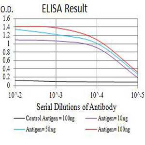
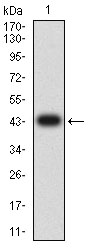
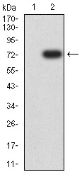
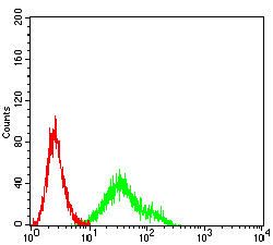
| WB, IHC, ICC, E |
|---|---|
| Primary Accession | Q08345 |
| Reactivity | Human |
| Host | Mouse |
| Clonality | Monoclonal |
| Clone Names | 4F2C12 |
| Isotype | Mouse IgG2b |
| Calculated MW | 101128 Da |
| Immunogen | Purified recombinant fragment of human CD167 (AA: extra 21-176) expressed in E. Coli. |
| Formulation | Purified antibody in PBS with 0.05% sodium azide |
| Gene ID | 780 |
|---|---|
| Other Names | DDR1;CAK; DDR; NEP; HGK2; PTK3; RTK6; TRKE; EDDR1; MCK10; NTRK4; PTK3A |
| Dilution | WB~~ 1/500 - 1/2000 IHC~~1:100~500 ICC~~N/A E~~ 1/10000 |
| Storage | Maintain refrigerated at 2-8°C for up to 6 months. For long term storage store at -20°C in small aliquots to prevent freeze-thaw cycles. |
| Precautions | CD167 is for research use only and not for use in diagnostic or therapeutic procedures. |
| Name | DDR1 |
|---|---|
| Synonyms | CAK, EDDR1, NEP, NTRK4, PTK3A, RTK6, TRK |
| Function | Tyrosine kinase that functions as a cell surface receptor for fibrillar collagen and regulates cell attachment to the extracellular matrix, remodeling of the extracellular matrix, cell migration, differentiation, survival and cell proliferation. Collagen binding triggers a signaling pathway that involves SRC and leads to the activation of MAP kinases. Regulates remodeling of the extracellular matrix by up-regulation of the matrix metalloproteinases MMP2, MMP7 and MMP9, and thereby facilitates cell migration and wound healing. Required for normal blastocyst implantation during pregnancy, for normal mammary gland differentiation and normal lactation. Required for normal ear morphology and normal hearing (By similarity). Promotes smooth muscle cell migration, and thereby contributes to arterial wound healing. Also plays a role in tumor cell invasion. Phosphorylates PTPN11. |
| Cellular Location | [Isoform 1]: Cell membrane; Single-pass type I membrane protein [Isoform 3]: Secreted. |
| Tissue Location | Detected in T-47D, MDA-MB-175 and HBL-100 breast carcinoma cells, A-431 epidermoid carcinoma cells, SW48 and SNU-C2B colon carcinoma cells and Hs 294T melanoma cells (at protein level) Expressed at low levels in most adult tissues and is highest in the brain, lung, placenta and kidney. Lower levels of expression are detected in melanocytes, heart, liver, skeletal muscle and pancreas Abundant in breast carcinoma cell lines. In the colonic mucosa, expressed in epithelia but not in the connective tissue of the lamina propria. In the thyroid gland, expressed in the epithelium of the thyroid follicles. In pancreas, expressed in the islets of Langerhans cells, but not in the surrounding epithelial cells of the exocrine pancreas. In kidney, expressed in the epithelia of the distal tubules Not expressed in connective tissue, endothelial cells, adipose tissue, muscle cells or cells of hematopoietic origin |
Research Areas
For Research Use Only. Not For Use In Diagnostic Procedures.
Application Protocols
Provided below are standard protocols that you may find useful for product applications.
REFERENCES
1.Am J Physiol Cell Physiol. 2015 May 1;308(9):C685-96.2.Med Oncol. 2013;30(3):626.
终于等到您。ABCEPTA(百远生物)抗体产品。
点击下方“我要评价 ”按钮提交您的反馈信息,您的反馈和评价是我们最宝贵的财富之一,
我们将在1-3个工作日内处理您的反馈信息。
如有疑问,联系:0512-88856768 tech-china@abcepta.com.
¥ 1,500.00
Cat# AO2709a


