


 癌症的基本特征包括细胞增殖、血管生成、迁移、凋亡逃避机制和细胞永生等。找到癌症发生过程中这些通路的关键标记物和对应的抗体用于检测至关重要。
 为您推荐一个泛素化位点预测神器——泛素化分析工具,可以为您的蛋白的泛素化位点作出预测和评分。
 细胞自噬受体图形绘图工具为你的蛋白的细胞受体结合位点作出预测和评分,识别结合到自噬通路中的蛋白是非常重要的,便于让我们理解自噬在正常生理、病理过程中的作用,如发育、细胞分化、神经退化性疾病、压力条件下、感染和癌症。
A4GALT Antibody (N-term)
Affinity Purified Rabbit Polyclonal Antibody (Pab)
- 产品详情
- 实验流程
- 背景知识
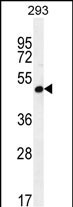
Application
| WB, E |
|---|---|
| Primary Accession | Q9NPC4 |
| Other Accession | NP_059132.1 |
| Reactivity | Human |
| Host | Rabbit |
| Clonality | Polyclonal |
| Isotype | Rabbit IgG |
| Calculated MW | 40499 Da |
| Antigen Region | 26-54 aa |
| Gene ID | 53947 |
|---|---|
| Other Names | Lactosylceramide 4-alpha-galactosyltransferase, Alpha-1, 4-N-acetylglucosaminyltransferase, Alpha-1, 4-galactosyltransferase, Alpha4Gal-T1, CD77 synthase, Globotriaosylceramide synthase, Gb3 synthase, P1/Pk synthase, UDP-galactose:beta-D-galactosyl-beta1-R 4-alpha-D-galactosyltransferase, A4GALT, A14GALT, A4GALT1 |
| Target/Specificity | This A4GALT antibody is generated from rabbits immunized with a KLH conjugated synthetic peptide between 26-54 amino acids from the N-terminal region of human A4GALT. |
| Dilution | WB~~1:1000 E~~Use at an assay dependent concentration. |
| Format | Purified polyclonal antibody supplied in PBS with 0.09% (W/V) sodium azide. This antibody is purified through a protein A column, followed by peptide affinity purification. |
| Storage | Maintain refrigerated at 2-8°C for up to 2 weeks. For long term storage store at -20°C in small aliquots to prevent freeze-thaw cycles. |
| Precautions | A4GALT Antibody (N-term) is for research use only and not for use in diagnostic or therapeutic procedures. |
| Name | A4GALT |
|---|---|
| Synonyms | A14GALT, A4GALT1 |
| Function | Glycosyltransferase involved in biosynthesis of P1, P(k) and the rare NOR antigens of P1PK histo-blood group system (PubMed:10747952, PubMed:10748143, PubMed:22965229, PubMed:26773500, PubMed:29572228, PubMed:33460651). Catalyzes the transfer of galactose from UDP-alpha-D-galactose in an alpha1,4 linkage to lactosylceramide/beta-D-galactosyl-(1->4)-beta-D-glucosyl-(1<->1)- ceramide(d18:1(4E)) to produce globotriaosylceramide/globoside Gb3Cer (d18:1(4E)), also known as P(k) antigen or CD77 (PubMed:10748143, PubMed:10747952, PubMed:26773500, PubMed:29572228, PubMed:22965229). Globoside Gb3Cer/P(k)/CD77 is a root structure in biosynthesis pathway of neutral glycosphingolipids of the globo-series. Broadly expressed in cell membranes of immune cells and other tissues, Gb3Cer/P(k)/CD77 antigen may participate in mechanisms that regulate cell-cell interactions and cell differentiation. In germinal center B cells, Gb3Cer/CD77 interacts with CD19 to drive centroblast-to-centrocyte transition, B cell receptor complex assembly and T follicular helper cell differentiation. In epithelium, Gb3Cer regulates CDH1/E-cadherin- dependent cell-cell adhesion counteracting epithelial-to-mesenchymal transition (By similarity) (PubMed:29572228). Synthesizes P1 glycan epitope (alpha-D-Gal-(1->4)-beta-D-Gal-(1->4)-GlcNAc-R) by transferring a galactose moiety to paragloboside nLc4 or to complex-type N-glycans (PubMed:26773500, PubMed:29572228, PubMed:33460651). Generates the rare NOR1 and NOR2 antigens which carry a Gal(alpha1->4)GalNAc terminal moiety and are both derived from Gb4Cer precursor (PubMed:22965229, PubMed:26773500, PubMed:33460651). Also able to transfer galactose to galactosylceramide/beta-D-Gal-(1<->1')-Cer (PubMed:10748143). |
| Cellular Location | Golgi apparatus membrane; Single- pass type II membrane protein |
| Tissue Location | Ubiquitous. Highly expressed in kidney, heart, spleen, liver, testis and placenta |
For Research Use Only. Not For Use In Diagnostic Procedures.
Provided below are standard protocols that you may find useful for product applications.
BACKGROUND
The protein encoded by this gene catalyzes the transfer of galactose to lactosylceramide to form globotriaosylceramide, which has been identified as the P(k) antigen of the P blood group system. The encoded protein, which is a type II membrane protein found in the Golgi, is also required for the synthesis of the bacterial verotoxins receptor.
REFERENCES
Zumbrun, S.D., et al. Infect. Immun. 78(11):4488-4499(2010)
Shin, I.S., et al. BMB Rep 42(5):310-314(2009)
Lund, N., et al. Blood 113(20):4980-4991(2009)
Okuda, T., et al. Glycobiology 18(12):1028-1035(2008)
Hellberg, A., et al. BMC Genet. 6, 49 (2005) :
终于等到您。ABCEPTA(百远生物)抗体产品。
点击下方“我要评价 ”按钮提交您的反馈信息,您的反馈和评价是我们最宝贵的财富之一,
我们将在1-3个工作日内处理您的反馈信息。
如有疑问,联系:0512-88856768 tech-china@abcepta.com.


