


 癌症的基本特征包括细胞增殖、血管生成、迁移、凋亡逃避机制和细胞永生等。找到癌症发生过程中这些通路的关键标记物和对应的抗体用于检测至关重要。
 为您推荐一个泛素化位点预测神器——泛素化分析工具,可以为您的蛋白的泛素化位点作出预测和评分。
 细胞自噬受体图形绘图工具为你的蛋白的细胞受体结合位点作出预测和评分,识别结合到自噬通路中的蛋白是非常重要的,便于让我们理解自噬在正常生理、病理过程中的作用,如发育、细胞分化、神经退化性疾病、压力条件下、感染和癌症。
IGF2BP2 Antibody (C-term)
Affinity Purified Rabbit Polyclonal Antibody (Pab)
- 产品详情
- 实验流程
- 背景知识
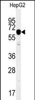
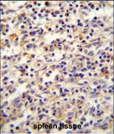
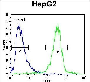
Application
| WB, IHC-P, FC, E |
|---|---|
| Primary Accession | Q9Y6M1 |
| Other Accession | Q5SF07, NP_001007226.1, NP_006539.3 |
| Reactivity | Human, Mouse |
| Predicted | Mouse |
| Host | Rabbit |
| Clonality | Polyclonal |
| Isotype | Rabbit IgG |
| Calculated MW | 66121 Da |
| Antigen Region | 530-556 aa |
| Gene ID | 10644 |
|---|---|
| Other Names | Insulin-like growth factor 2 mRNA-binding protein 2, IGF2 mRNA-binding protein 2, IMP-2, Hepatocellular carcinoma autoantigen p62, IGF-II mRNA-binding protein 2, VICKZ family member 2, IGF2BP2, IMP2, VICKZ2 |
| Target/Specificity | This IGF2BP2 antibody is generated from rabbits immunized with a KLH conjugated synthetic peptide between 530-556 amino acids from the C-terminal region of human IGF2BP2. |
| Dilution | WB~~1:1000 IHC-P~~1:100~500 FC~~1:10~50 E~~Use at an assay dependent concentration. |
| Format | Purified polyclonal antibody supplied in PBS with 0.09% (W/V) sodium azide. This antibody is purified through a protein A column, followed by peptide affinity purification. |
| Storage | Maintain refrigerated at 2-8°C for up to 2 weeks. For long term storage store at -20°C in small aliquots to prevent freeze-thaw cycles. |
| Precautions | IGF2BP2 Antibody (C-term) is for research use only and not for use in diagnostic or therapeutic procedures. |
| Name | IGF2BP2 |
|---|---|
| Synonyms | IMP2, VICKZ2 |
| Function | RNA-binding factor that recruits target transcripts to cytoplasmic protein-RNA complexes (mRNPs). This transcript 'caging' into mRNPs allows mRNA transport and transient storage. It also modulates the rate and location at which target transcripts encounter the translational apparatus and shields them from endonuclease attacks or microRNA-mediated degradation (By similarity). Preferentially binds to N6-methyladenosine (m6A)-containing mRNAs and increases their stability (PubMed:29476152). Binds to the 5'-UTR of the insulin-like growth factor 2 (IGF2) mRNAs (PubMed:9891060). Binding is isoform- specific. Binds to beta-actin/ACTB and MYC transcripts. Increases MYC mRNA stability by binding to the coding region instability determinant (CRD) and binding is enhanced by m6A-modification of the CRD (PubMed:29476152). |
| Cellular Location | Nucleus. Cytoplasm. Cytoplasm, P-body. Cytoplasm, Stress granule. Note=Localized in cytoplasmic mRNP granules containing untranslated mRNAs. Localizes at the connecting piece and the tail of the spermatozoa. In response to cellular stress, such as oxidative stress, recruited to stress granules |
| Tissue Location | Expressed in oocytes, granulosa cells of small and growing follicles, Leydig cells, spermatogonia and semen (at protein level). Expressed in testicular cancer (at protein level). Expressed weakly in heart, placenta, skeletal muscle, bone marrow, colon, kidney, salivary glands, testis and pancreas. Detected in fetal liver, fetal ovary, gonocytes and interstitial cells of the testis |
For Research Use Only. Not For Use In Diagnostic Procedures.
Provided below are standard protocols that you may find useful for product applications.
BACKGROUND
This gene encodes a member of the IGF-II mRNA-binding protein (IMP) family. The protein encoded by this gene contains several four KH domains and two RRM domains. It functions by binding to the 5' UTR of the insulin-like growth factor 2 (IGF2) mRNA and regulating IGF2 translation. Alternate transcriptional splice variants, encoding different isoforms, have been characterized.
REFERENCES
Bailey, S.D., et al. Diabetes Care 33(10):2250-2253(2010)
Pechlivanis, S., et al. Arterioscler. Thromb. Vasc. Biol. 30(9):1867-1872(2010)
Heni, M., et al. Diabetes (2010) In press :
Rodriguez, S., et al. Growth Horm. IGF Res. 20(4):310-318(2010)
Voight, B.F., et al. Nat. Genet. 42(7):579-589(2010)
终于等到您。ABCEPTA(百远生物)抗体产品。
点击下方“我要评价 ”按钮提交您的反馈信息,您的反馈和评价是我们最宝贵的财富之一,
我们将在1-3个工作日内处理您的反馈信息。
如有疑问,联系:0512-88856768 tech-china@abcepta.com.


