


 癌症的基本特征包括细胞增殖、血管生成、迁移、凋亡逃避机制和细胞永生等。找到癌症发生过程中这些通路的关键标记物和对应的抗体用于检测至关重要。
 为您推荐一个泛素化位点预测神器——泛素化分析工具,可以为您的蛋白的泛素化位点作出预测和评分。
 细胞自噬受体图形绘图工具为你的蛋白的细胞受体结合位点作出预测和评分,识别结合到自噬通路中的蛋白是非常重要的,便于让我们理解自噬在正常生理、病理过程中的作用,如发育、细胞分化、神经退化性疾病、压力条件下、感染和癌症。
CXCR3 Antibody (Center)
Affinity Purified Rabbit Polyclonal Antibody (Pab)
- 产品详情
- 实验流程
- 背景知识
Application
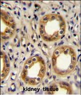
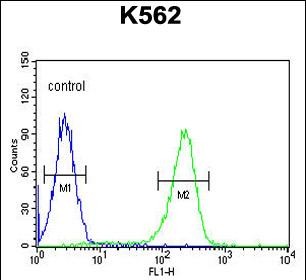
| WB, IHC-P, FC, E |
|---|---|
| Primary Accession | P49682 |
| Other Accession | NP_001495.1, NP_001136269.1 |
| Reactivity | Human |
| Host | Rabbit |
| Clonality | Polyclonal |
| Isotype | Rabbit IgG |
| Calculated MW | 40660 Da |
| Antigen Region | 140-167 aa |
| Gene ID | 2833 |
|---|---|
| Other Names | C-X-C chemokine receptor type 3, CXC-R3, CXCR-3, CKR-L2, G protein-coupled receptor 9, Interferon-inducible protein 10 receptor, IP-10 receptor, CD183, CXCR3, GPR9 |
| Target/Specificity | This CXCR3 antibody is generated from rabbits immunized with a KLH conjugated synthetic peptide between 140-167 amino acids of human CXCR3. |
| Dilution | WB~~1:1000 IHC-P~~1:100~500 FC~~1:10~50 E~~Use at an assay dependent concentration. |
| Format | Purified polyclonal antibody supplied in PBS with 0.05% (V/V) Proclin 300. This antibody is prepared by Saturated Ammonium Sulfate (SAS) precipitation followed by dialysis against PBS. |
| Storage | Maintain refrigerated at 2-8°C for up to 2 weeks. For long term storage store at -20°C in small aliquots to prevent freeze-thaw cycles. |
| Precautions | CXCR3 Antibody (Center) is for research use only and not for use in diagnostic or therapeutic procedures. |
| Name | CXCR3 |
|---|---|
| Synonyms | GPR9 |
| Function | [Isoform 1]: Receptor for the C-X-C chemokine CXCL9, CXCL10 and CXCL11 and mediates the proliferation, survival and angiogenic activity of human mesangial cells (HMC) through a heterotrimeric G- protein signaling pathway (PubMed:12782716). Binds to CCL21. Probably promotes cell chemotaxis response. Upon activation by PF4, induces activated T-lymphocytes migration mediated via downstream Ras/extracellular signal-regulated kinase (ERK) signaling. [Isoform 3]: Mediates the activity of CXCL11. |
| Cellular Location | [Isoform 1]: Cell membrane; Multi-pass membrane protein |
| Tissue Location | Isoform 1 and isoform 2 are mainly expressed in heart, kidney, liver and skeletal muscle. Isoform 1 is also expressed in placenta. Isoform 2 is expressed in endothelial cells. Expressed in T-cells (at protein level). |
For Research Use Only. Not For Use In Diagnostic Procedures.
Provided below are standard protocols that you may find useful for product applications.
BACKGROUND
This gene encodes a G protein-coupled receptor with selectivity for three chemokines, termed IP10 (interferon-g-inducible 10 kDa protein), Mig (monokine induced by interferon-g) and I-TAC (interferon-inducible T cell a-chemoattractant). IP10, Mig and I-TAC belong to the structural subfamily of CXC chemokines, in which a single amino acid residue separates the first two of four highly conserved Cys residues. Binding of chemokines to this protein induces cellular responses that are involved in leukocyte traffic, most notably integrin activation, cytoskeletal changes and chemotactic migration. Inhibition by Bordetella pertussis toxin suggests that heterotrimeric G protein of the Gi-subclass couple to this protein. Signal transduction has not been further analyzed but may include the same enzymes that were identified in the signaling cascade induced by other chemokine receptors. As a consequence of chemokine-induced cellular desensitization (phosphorylation-dependent receptor internalization), cellular responses are typically rapid and short in duration. Cellular responsiveness is restored after dephosphorylation of intracellular receptors and subsequent recycling to the cell surface. This gene is prominently expressed in in vitro cultured effector/memory T cells, and in T cells present in many types of inflamed tissues. In addition, IP10, Mig and I-TAC are commonly produced by local cells in inflammatory lesion, suggesting that this gene and its chemokines participate in the recruitment of inflammatory cells. Therefore, this protein is a target for the development of small molecular weight antagonists, which may be used in the treatment of diverse inflammatory diseases. Multiple transcript variants encoding different isoforms have been found for this gene.
REFERENCES
Zhou, J., et al. J. Exp. Med. 207(9):1951-1966(2010)
Wang, Y., et al. J. Hum. Genet. 55(8):490-494(2010)
Schuurhof, A., et al. Pediatr. Pulmonol. 45(6):608-613(2010)
Miekus, K., et al. Folia Histochem. Cytobiol. 48(1):104-111(2010)
Ohri, C.M., et al. BMC Cancer 10, 172 (2010) :
终于等到您。ABCEPTA(百远生物)抗体产品。
点击下方“我要评价 ”按钮提交您的反馈信息,您的反馈和评价是我们最宝贵的财富之一,
我们将在1-3个工作日内处理您的反馈信息。
如有疑问,联系:0512-88856768 tech-china@abcepta.com.


