


 癌症的基本特征包括细胞增殖、血管生成、迁移、凋亡逃避机制和细胞永生等。找到癌症发生过程中这些通路的关键标记物和对应的抗体用于检测至关重要。
 为您推荐一个泛素化位点预测神器——泛素化分析工具,可以为您的蛋白的泛素化位点作出预测和评分。
 细胞自噬受体图形绘图工具为你的蛋白的细胞受体结合位点作出预测和评分,识别结合到自噬通路中的蛋白是非常重要的,便于让我们理解自噬在正常生理、病理过程中的作用,如发育、细胞分化、神经退化性疾病、压力条件下、感染和癌症。
FOLR1 Antibody (N-term)
Affinity Purified Rabbit Polyclonal Antibody (Pab)

- 产品详情
- 实验流程
- 背景知识
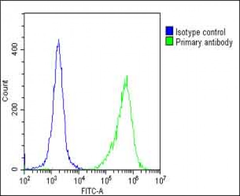
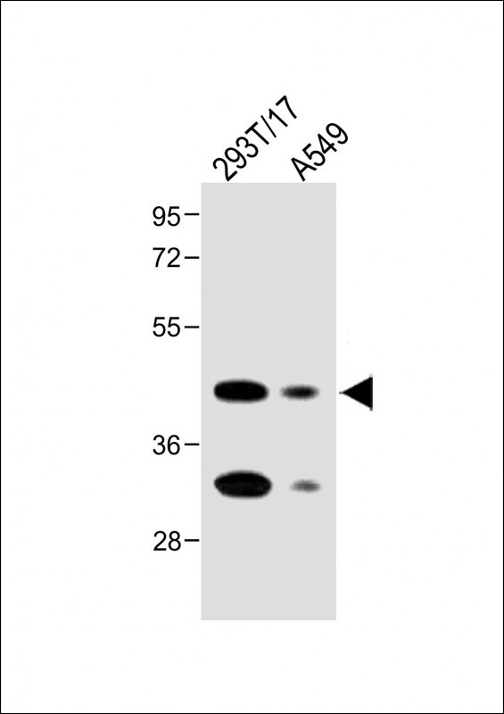
Application
| FC, WB, IHC-P-Leica, E |
|---|---|
| Primary Accession | P15328 |
| Other Accession | O15254, NP_000793.1 |
| Reactivity | Human, Mouse |
| Host | Rabbit |
| Clonality | Polyclonal |
| Isotype | Rabbit IgG |
| Calculated MW | 29819 Da |
| Antigen Region | 33-68 aa |
| Gene ID | 2348 |
|---|---|
| Other Names | Folate receptor alpha, FR-alpha, Adult folate-binding protein, FBP, Folate receptor 1, Folate receptor, adult, KB cells FBP, Ovarian tumor-associated antigen MOv18, FOLR1, FOLR |
| Target/Specificity | This FOLR1 antibody is generated from rabbits immunized with a KLH conjugated synthetic peptide between 33-68 amino acids from the N-terminal region of human FOLR1. |
| Dilution | FC~~1:25 WB~~1:2000 IHC-P-Leica~~1:2000 E~~Use at an assay dependent concentration. |
| Format | Purified polyclonal antibody supplied in PBS with 0.09% (W/V) sodium azide. This antibody is purified through a protein A column, followed by peptide affinity purification. |
| Storage | Maintain refrigerated at 2-8°C for up to 2 weeks. For long term storage store at -20°C in small aliquots to prevent freeze-thaw cycles. |
| Precautions | FOLR1 Antibody (N-term) is for research use only and not for use in diagnostic or therapeutic procedures. |
| Name | FOLR1 |
|---|---|
| Synonyms | FOLR |
| Function | Binds to folate and reduced folic acid derivatives and mediates delivery of 5-methyltetrahydrofolate and folate analogs into the interior of cells (PubMed:19074442, PubMed:23851396, PubMed:23934049, PubMed:2527252, PubMed:8033114, PubMed:8567728). Has high affinity for folate and folic acid analogs at neutral pH (PubMed:23851396, PubMed:23934049, PubMed:2527252, PubMed:8033114, PubMed:8567728). Exposure to slightly acidic pH after receptor endocytosis triggers a conformation change that strongly reduces its affinity for folates and mediates their release (PubMed:8567728). Required for normal embryonic development and normal cell proliferation (By similarity). |
| Cellular Location | Cell membrane; Lipid-anchor, GPI-anchor Apical cell membrane; Lipid-anchor, GPI- anchor Basolateral cell membrane; Lipid-anchor, GPI-like-anchor. Secreted Cytoplasmic vesicle. Cytoplasmic vesicle, clathrin-coated vesicle. Endosome. Note=Endocytosed into cytoplasmic vesicles and then recycled to the cell membrane |
| Tissue Location | Primarily expressed in tissues of epithelial origin. Expression is increased in malignant tissues. Expressed in kidney, lung and cerebellum. Detected in placenta and thymus epithelium. |
For Research Use Only. Not For Use In Diagnostic Procedures.
Provided below are standard protocols that you may find useful for product applications.
BACKGROUND
The protein encoded by this gene is a member of the folate receptor family. Members of this gene family bind folic acid and its reduced derivatives, and transport 5-methyltetrahydrofolate into cells. This gene product is a secreted protein that either anchors to membranes via a glycosyl-phosphatidylinositol linkage or exists in a soluble form. Mutations in this gene have been associated with neurodegeneration due to cerebral folate transport deficiency. Due to the presence of two promoters, multiple transcription start sites, and alternative splicing, multiple transcript variants encoding the same protein have been found for this gene.
REFERENCES
Sivakumaran, S., et al. J. Steroid Biochem. Mol. Biol. 122(5):333-340(2010)
Bailey, S.D., et al. Diabetes Care 33(10):2250-2253(2010)
O'Byrne, M.R., et al. Birth Defects Res. Part A Clin. Mol. Teratol. 88(8):689-694(2010)
Jugessur, A., et al. PLoS ONE 5 (7), E11493 (2010) :
Elwood, P.C., et al. Biochemistry 36(6):1467-1478(1997)
终于等到您。ABCEPTA(百远生物)抗体产品。
点击下方“我要评价 ”按钮提交您的反馈信息,您的反馈和评价是我们最宝贵的财富之一,
我们将在1-3个工作日内处理您的反馈信息。
如有疑问,联系:0512-88856768 tech-china@abcepta.com.


