


 癌症的基本特征包括细胞增殖、血管生成、迁移、凋亡逃避机制和细胞永生等。找到癌症发生过程中这些通路的关键标记物和对应的抗体用于检测至关重要。
 为您推荐一个泛素化位点预测神器——泛素化分析工具,可以为您的蛋白的泛素化位点作出预测和评分。
 细胞自噬受体图形绘图工具为你的蛋白的细胞受体结合位点作出预测和评分,识别结合到自噬通路中的蛋白是非常重要的,便于让我们理解自噬在正常生理、病理过程中的作用,如发育、细胞分化、神经退化性疾病、压力条件下、感染和癌症。
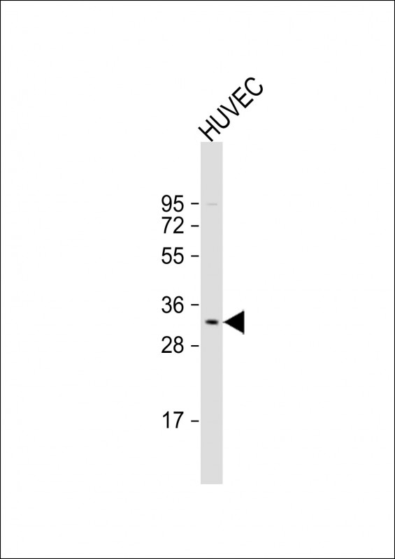
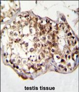
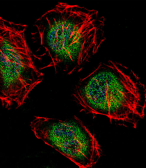
CAF-1 Antibody (N-term)
Affinity Purified Rabbit Polyclonal Antibody (Pab)
- 产品详情
- 实验流程
- 背景知识
Application
| IHC-P, IF, WB, E |
|---|---|
| Primary Accession | Q9UIV1 |
| Other Accession | Q3KQ85, Q60809, Q08BM8, Q5ZJV9, Q3ZC01, NP_473367.2 |
| Reactivity | Human |
| Predicted | Bovine, Chicken, Zebrafish, Mouse, Xenopus |
| Host | Rabbit |
| Clonality | Polyclonal |
| Isotype | Rabbit IgG |
| Calculated MW | 32745 Da |
| Antigen Region | 34-61 aa |
| Gene ID | 29883 |
|---|---|
| Other Names | CCR4-NOT transcription complex subunit 7, BTG1-binding factor 1, CCR4-associated factor 1, CAF-1, Caf1a, CNOT7, CAF1 |
| Target/Specificity | This CAF-1 antibody is generated from rabbits immunized with a KLH conjugated synthetic peptide between 34-61 amino acids from the N-terminal region of human CAF-1. |
| Dilution | IHC-P~~1:100~500 IF~~1:10~50 WB~~1:1000 E~~Use at an assay dependent concentration. |
| Format | Purified polyclonal antibody supplied in PBS with 0.09% (W/V) sodium azide. This antibody is purified through a protein A column, followed by peptide affinity purification. |
| Storage | Maintain refrigerated at 2-8°C for up to 2 weeks. For long term storage store at -20°C in small aliquots to prevent freeze-thaw cycles. |
| Precautions | CAF-1 Antibody (N-term) is for research use only and not for use in diagnostic or therapeutic procedures. |
| Name | CNOT7 |
|---|---|
| Synonyms | CAF1 |
| Function | Has 3'-5' poly(A) exoribonuclease activity for synthetic poly(A) RNA substrate (PubMed:19276069, PubMed:20634287, PubMed:31439799). Its function seems to be partially redundant with that of CNOT8 (PubMed:19605561). Catalytic component of the CCR4-NOT complex which is one of the major cellular mRNA deadenylases and is linked to various cellular processes including bulk mRNA degradation, miRNA-mediated repression, translational repression during translational initiation and general transcription regulation (PubMed:19276069, PubMed:20634287, PubMed:31439799). During miRNA- mediated repression the complex also seems to act as translational repressor during translational initiation (PubMed:20065043). Additional complex functions may be a consequence of its influence on mRNA expression (PubMed:19276069, PubMed:23236473). Associates with members of the BTG family such as TOB1 and BTG2 and is required for their anti- proliferative activity (PubMed:19276069, PubMed:23236473). |
| Cellular Location | Nucleus. Cytoplasm, P-body {ECO:0000250|UniProtKB:Q60809}. Cytoplasm, Cytoplasmic ribonucleoprotein granule. Note=NANOS2 promotes its localization to P-body (By similarity). Recruited to cytoplasmic ribonucleoprotein membraneless compartments by CAPRIN1, promoting deadenylation of mRNAs (PubMed:31439799) {ECO:0000250|UniProtKB:Q60809, ECO:0000269|PubMed:31439799} |
For Research Use Only. Not For Use In Diagnostic Procedures.
Provided below are standard protocols that you may find useful for product applications.
BACKGROUND
The protein encoded by this gene binds to an anti-proliferative protein, B-cell translocation protein 1, which negatively regulates cell proliferation. Binding of the two proteins, which is driven by phosphorylation of the anti-proliferative protein, causes signaling events in cell division that lead to changes in cell proliferation associated with cell-cell contact. The protein has both mouse and yeast orthologs. Alternate splicing of this gene results in two transcript variants encoding different isoforms.
REFERENCES
Lau, N.C., et al. Biochem. J. 422(3):443-453(2009)
Aslam, A., et al. Mol. Biol. Cell 20(17):3840-3850(2009)
Miyasaka, T., et al. Cancer Sci. 99(4):755-761(2008)
Nishida, K., et al. Acta Crystallogr. Sect. F Struct. Biol. Cryst. Commun. 63 (PT 12), 1061-1063 (2007) :
Robin-Lespinasse, Y., et al. J. Cell. Sci. 120 (PT 4), 638-647 (2007) :
终于等到您。ABCEPTA(百远生物)抗体产品。
点击下方“我要评价 ”按钮提交您的反馈信息,您的反馈和评价是我们最宝贵的财富之一,
我们将在1-3个工作日内处理您的反馈信息。
如有疑问,联系:0512-88856768 tech-china@abcepta.com.


