


 癌症的基本特征包括细胞增殖、血管生成、迁移、凋亡逃避机制和细胞永生等。找到癌症发生过程中这些通路的关键标记物和对应的抗体用于检测至关重要。
 为您推荐一个泛素化位点预测神器——泛素化分析工具,可以为您的蛋白的泛素化位点作出预测和评分。
 细胞自噬受体图形绘图工具为你的蛋白的细胞受体结合位点作出预测和评分,识别结合到自噬通路中的蛋白是非常重要的,便于让我们理解自噬在正常生理、病理过程中的作用,如发育、细胞分化、神经退化性疾病、压力条件下、感染和癌症。
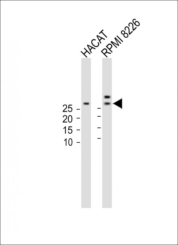
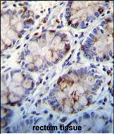
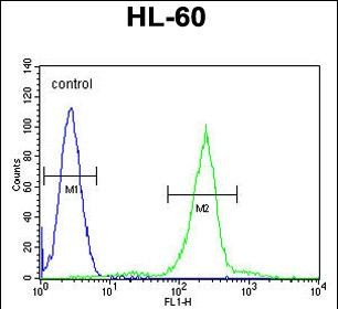
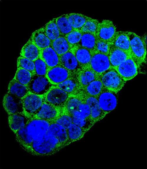
C1QC Antibody (Center)
Affinity Purified Rabbit Polyclonal Antibody (Pab)
- 产品详情
- 实验流程
- 背景知识
Application
| WB, IHC-P, FC, IF, E |
|---|---|
| Primary Accession | P02747 |
| Other Accession | NP_758957.2 |
| Reactivity | Human |
| Host | Rabbit |
| Clonality | Polyclonal |
| Isotype | Rabbit IgG |
| Calculated MW | 25774 Da |
| Antigen Region | 93-120 aa |
| Gene ID | 714 |
|---|---|
| Other Names | Complement C1q subcomponent subunit C, C1QC, C1QG |
| Target/Specificity | This C1QC antibody is generated from rabbits immunized with a KLH conjugated synthetic peptide between 93-120 amino acids from the Central region of human C1QC. |
| Dilution | WB~~1:1000 IHC-P~~1:100~500 FC~~1:10~50 IF~~1:10~50 E~~Use at an assay dependent concentration. |
| Format | Purified polyclonal antibody supplied in PBS with 0.05% (V/V) Proclin 300. This antibody is prepared by Saturated Ammonium Sulfate (SAS) precipitation followed by dialysis against PBS. |
| Storage | Maintain refrigerated at 2-8°C for up to 2 weeks. For long term storage store at -20°C in small aliquots to prevent freeze-thaw cycles. |
| Precautions | C1QC Antibody (Center) is for research use only and not for use in diagnostic or therapeutic procedures. |
| Name | C1QC {ECO:0000303|PubMed:1706597, ECO:0000312|HGNC:HGNC:1245} |
|---|---|
| Function | Core component of the complement C1 complex, a multiprotein complex that initiates the classical pathway of the complement system, a cascade of proteins that leads to phagocytosis and breakdown of pathogens and signaling that strengthens the adaptive immune system (PubMed:12847249, PubMed:19006321, PubMed:24626930, PubMed:29449492, PubMed:3258649, PubMed:34155115, PubMed:6249812, PubMed:6776418). The classical complement pathway is initiated by the C1Q subcomplex of the C1 complex, which specifically binds IgG or IgM immunoglobulins complexed with antigens, forming antigen-antibody complexes on the surface of pathogens: C1QA, together with C1QB and C1QC, specifically recognizes and binds the Fc regions of IgG or IgM via its C1q domain (PubMed:12847249, PubMed:19006321, PubMed:24626930, PubMed:29449492, PubMed:3258649, PubMed:6776418). Immunoglobulin-binding activates the proenzyme C1R, which cleaves C1S, initiating the proteolytic cascade of the complement system (PubMed:29449492). The C1Q subcomplex is activated by a hexamer of IgG complexed with antigens, while it is activated by a pentameric IgM (PubMed:19706439, PubMed:24626930, PubMed:29449492). The C1Q subcomplex also recognizes and binds phosphatidylserine exposed on the surface of cells undergoing programmed cell death, possibly promoting activation of the complement system (PubMed:18250442). |
| Cellular Location | Secreted. Cell surface. Note=Specifically binds IgG or IgM immunoglobulins complexed with antigens, forming antigen-antibody complexes on the surface of pathogens. |
For Research Use Only. Not For Use In Diagnostic Procedures.
Provided below are standard protocols that you may find useful for product applications.
BACKGROUND
This gene encodes a major constituent of the human complement subcomponent C1q. C1q associates with C1r and C1s in order to yield the first component of the serum complement system. A deficiency in C1q has been associated with lupus erythematosus and glomerulonephritis. C1q is composed of 18 polypeptide chains: six A-chains, six B-chains, and six C-chains. Each chain contains a collagen-like region located near the N-terminus, and a C-terminal globular region. The A-, B-, and C-chains are arranged in the order A-C-B on chromosome 1. This gene encodes the C-chain polypeptide of human complement subcomponent C1q. Alternatively spliced transcript variants that encode the same protein have been found for this gene.
REFERENCES
Fraser, D.A., et al. J. Immunol. 185(7):3932-3939(2010)
Bailey, S.D., et al. Diabetes Care 33(10):2250-2253(2010)
Rafiq, S., et al. Clin. Exp. Immunol. 161(2):284-289(2010)
Han, S., et al. Hum. Immunol. 71(7):727-730(2010)
Rajaraman, P., et al. Cancer Epidemiol. Biomarkers Prev. 19(5):1356-1361(2010)
终于等到您。ABCEPTA(百远生物)抗体产品。
点击下方“我要评价 ”按钮提交您的反馈信息,您的反馈和评价是我们最宝贵的财富之一,
我们将在1-3个工作日内处理您的反馈信息。
如有疑问,联系:0512-88856768 tech-china@abcepta.com.


