


 癌症的基本特征包括细胞增殖、血管生成、迁移、凋亡逃避机制和细胞永生等。找到癌症发生过程中这些通路的关键标记物和对应的抗体用于检测至关重要。
 为您推荐一个泛素化位点预测神器——泛素化分析工具,可以为您的蛋白的泛素化位点作出预测和评分。
 细胞自噬受体图形绘图工具为你的蛋白的细胞受体结合位点作出预测和评分,识别结合到自噬通路中的蛋白是非常重要的,便于让我们理解自噬在正常生理、病理过程中的作用,如发育、细胞分化、神经退化性疾病、压力条件下、感染和癌症。
AXIN1 Antibody (C-term)
Affinity Purified Rabbit Polyclonal Antibody (Pab)
- 产品详情
- 实验流程
- 背景知识
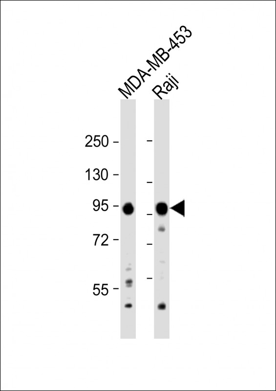
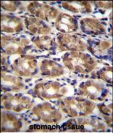
Application
| WB, IHC-P, E |
|---|---|
| Primary Accession | O15169 |
| Other Accession | NP_851393.1 |
| Reactivity | Human, Mouse |
| Host | Rabbit |
| Clonality | Polyclonal |
| Isotype | Rabbit IgG |
| Calculated MW | 95635 Da |
| Antigen Region | 710-738 aa |
| Gene ID | 8312 |
|---|---|
| Other Names | Axin-1, Axis inhibition protein 1, hAxin, AXIN1, AXIN |
| Target/Specificity | This AXIN1 antibody is generated from rabbits immunized with a KLH conjugated synthetic peptide between 710-738 amino acids from the C-terminal region of human AXIN1. |
| Dilution | WB~~1:1000 IHC-P~~1:100~500 E~~Use at an assay dependent concentration. |
| Format | Purified polyclonal antibody supplied in PBS with 0.09% (W/V) sodium azide. This antibody is purified through a protein A column, followed by peptide affinity purification. |
| Storage | Maintain refrigerated at 2-8°C for up to 2 weeks. For long term storage store at -20°C in small aliquots to prevent freeze-thaw cycles. |
| Precautions | AXIN1 Antibody (C-term) is for research use only and not for use in diagnostic or therapeutic procedures. |
| Name | AXIN1 |
|---|---|
| Synonyms | AXIN |
| Function | Component of the beta-catenin destruction complex required for regulating CTNNB1 levels through phosphorylation and ubiquitination, and modulating Wnt-signaling (PubMed:12192039, PubMed:27098453, PubMed:28829046). Controls dorsoventral patterning via two opposing effects; down-regulates CTNNB1 to inhibit the Wnt signaling pathway and ventralize embryos, but also dorsalizes embryos by activating a Wnt-independent JNK signaling pathway (PubMed:12192039). In Wnt signaling, probably facilitates the phosphorylation of CTNNB1 and APC by GSK3B (PubMed:12192039). Likely to function as a tumor suppressor. Enhances TGF-beta signaling by recruiting the RNF111 E3 ubiquitin ligase and promoting the degradation of inhibitory SMAD7 (PubMed:16601693). Also a component of the AXIN1- HIPK2-TP53 complex which controls cell growth, apoptosis and development (PubMed:17210684). Facilitates the phosphorylation of TP53 by HIPK2 upon ultraviolet irradiation (PubMed:17210684). |
| Cellular Location | Cytoplasm. Nucleus. Membrane {ECO:0000250|UniProtKB:O35625} Cell membrane {ECO:0000250|UniProtKB:O35625}. Note=MACF1 is required for its translocation to cell membrane (By similarity). On UV irradiation, translocates to the nucleus and colocalizes with DAAX (PubMed:17210684). {ECO:0000250|UniProtKB:O35625, ECO:0000269|PubMed:17210684} |
| Tissue Location | Ubiquitously expressed. |
For Research Use Only. Not For Use In Diagnostic Procedures.
Provided below are standard protocols that you may find useful for product applications.
BACKGROUND
This gene encodes a cytoplasmic protein which contains a regulation of G-protein signaling (RGS) domain and a dishevelled and axin (DIX) domain. The encoded protein interacts with adenomatosis polyposis coli, catenin beta-1, glycogen synthase kinase 3 beta, protein phosphate 2, and itself. This protein functions as a negative regulator of the wingless-type MMTV integration site family, member 1 (WNT) signaling pathway and can induce apoptosis. The crystal structure of a portion of this protein, alone and in a complex with other proteins, has been resolved. Mutations in this gene have been associated with hepatocellular carcinoma, hepatoblastomas, ovarian endometriod adenocarcinomas, and medullablastomas. Two transcript variants encoding distinct isoforms have been identified for this gene.
REFERENCES
Sue Ng, S., et al. Biol. Chem. 391 (2-3), 171-180 (2010) :
Yang, L.H., et al. Mol. Cancer 9, 25 (2010) :
Wooten, E.C., et al. PLoS ONE 5 (1), E8830 (2010) :
Kameoka, M., et al. AIDS Res. Hum. Retroviruses 25(10):1005-1011(2009)
Li, Q., et al. Nat. Cell Biol. 11(9):1128-1134(2009)
终于等到您。ABCEPTA(百远生物)抗体产品。
点击下方“我要评价 ”按钮提交您的反馈信息,您的反馈和评价是我们最宝贵的财富之一,
我们将在1-3个工作日内处理您的反馈信息。
如有疑问,联系:0512-88856768 tech-china@abcepta.com.


