


 癌症的基本特征包括细胞增殖、血管生成、迁移、凋亡逃避机制和细胞永生等。找到癌症发生过程中这些通路的关键标记物和对应的抗体用于检测至关重要。
 为您推荐一个泛素化位点预测神器——泛素化分析工具,可以为您的蛋白的泛素化位点作出预测和评分。
 细胞自噬受体图形绘图工具为你的蛋白的细胞受体结合位点作出预测和评分,识别结合到自噬通路中的蛋白是非常重要的,便于让我们理解自噬在正常生理、病理过程中的作用,如发育、细胞分化、神经退化性疾病、压力条件下、感染和癌症。
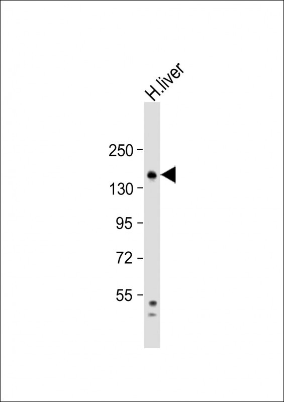
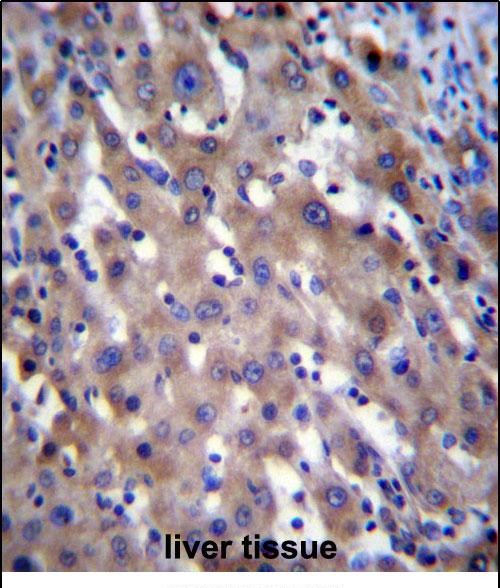
RPGR Antibody (C-term)
Affinity Purified Rabbit Polyclonal Antibody (Pab)
- 产品详情
- 实验流程
- 背景知识
Application
| WB, IHC-P, E |
|---|---|
| Primary Accession | Q92834 |
| Other Accession | NP_000319.1 |
| Reactivity | Human |
| Host | Rabbit |
| Clonality | Polyclonal |
| Isotype | Rabbit IgG |
| Calculated MW | 113387 Da |
| Antigen Region | 744-772 aa |
| Gene ID | 6103 |
|---|---|
| Other Names | X-linked retinitis pigmentosa GTPase regulator, RPGR, RP3, XLRP3 |
| Target/Specificity | This RPGR antibody is generated from rabbits immunized with a KLH conjugated synthetic peptide between 744-772 amino acids of human RPGR. |
| Dilution | WB~~1:1000 IHC-P~~1:100~500 E~~Use at an assay dependent concentration. |
| Format | Purified polyclonal antibody supplied in PBS with 0.09% (W/V) sodium azide. This antibody is purified through a protein A column, followed by peptide affinity purification. |
| Storage | Maintain refrigerated at 2-8°C for up to 2 weeks. For long term storage store at -20°C in small aliquots to prevent freeze-thaw cycles. |
| Precautions | RPGR Antibody (C-term) is for research use only and not for use in diagnostic or therapeutic procedures. |
| Name | RPGR (HGNC:10295) |
|---|---|
| Synonyms | RP3, XLRP3 |
| Function | Acts as a guanine-nucleotide releasing factor (GEF) for RAB8A and RAB37 by promoting the conversion of inactive RAB-GDP to the active form RAB-GTP (PubMed:20631154). GEF activity towards RAB8A may facilitate ciliary trafficking by modulating ciliary intracellular localization of RAB8A (PubMed:20631154). GEF activity towards RAB37 maintains autophagic homeostasis and retinal function (By similarity). Involved in photoreceptor integrity (By similarity). May control cilia formation by regulating actin stress filaments and cell contractility. May be involved in microtubule organization and regulation of transport in primary cilia (PubMed:21933838). May play a critical role in spermatogenesis and in intraflagellar transport processes (By similarity). |
| Cellular Location | Cytoplasm, cytoskeleton, flagellum axoneme {ECO:0000250|UniProtKB:Q9R0X5}. Golgi apparatus. Cell projection, cilium {ECO:0000250|UniProtKB:Q9R0X5}. Note=In the retinal photoreceptor cell layer, localizes at the connecting cilium (By similarity). Colocalizes with WHRN in the photoreceptor connecting cilium (By similarity) Colocalizes with CEP290 in the photoreceptor connecting cilium (By similarity). Colocalizes with RPGRIP1 in the photoreceptor connecting cilium (By similarity). Colocalizes with RPGR at the primary cilia of epithelial cells (By similarity). {ECO:0000250|UniProtKB:Q9N1T2, ECO:0000250|UniProtKB:Q9R0X5} |
| Tissue Location | Heart, brain, placenta, lung, liver, muscle, kidney, retina, pancreas and fetal retinal pigment epithelium. Isoform 3 is found only in the retina. Colocalizes with RPGRIP1 in the outer segment of rod photoreceptors and cone outer segments |
For Research Use Only. Not For Use In Diagnostic Procedures.
Provided below are standard protocols that you may find useful for product applications.
BACKGROUND
This gene encodes a protein with a series of six RCC1-like domains (RLDs), characteristic of the highly conserved guanine nucleotide exchange factors. The encoded protein is found in the Golgi body and interacts with RPGRIP1. This protein localizes to the outer segment of rod photoreceptors and is essential for their viability. Mutations in this gene have been associated with X-linked retinitis pigmentosa (XLRP). Multiple alternatively spliced transcript variants that encode different isoforms of this gene have been reported, but the full-length natures of only some have been determined.
REFERENCES
Clark, G.R., et al. Ophthalmology 117(11):2169-2177(2010)
Schmid, F., et al. Invest. Ophthalmol. Vis. Sci. 51(3):1628-1635(2010)
Ji, Y., et al. Curr. Eye Res. 35(1):73-79(2010)
Sheng, X., et al. Mol. Vis. 16, 1620-1628 (2010) :
Murga-Zamalloa, C.A., et al. J. Genet. 88(4):399-407(2009)
终于等到您。ABCEPTA(百远生物)抗体产品。
点击下方“我要评价 ”按钮提交您的反馈信息,您的反馈和评价是我们最宝贵的财富之一,
我们将在1-3个工作日内处理您的反馈信息。
如有疑问,联系:0512-88856768 tech-china@abcepta.com.


