


 癌症的基本特征包括细胞增殖、血管生成、迁移、凋亡逃避机制和细胞永生等。找到癌症发生过程中这些通路的关键标记物和对应的抗体用于检测至关重要。
 为您推荐一个泛素化位点预测神器——泛素化分析工具,可以为您的蛋白的泛素化位点作出预测和评分。
 细胞自噬受体图形绘图工具为你的蛋白的细胞受体结合位点作出预测和评分,识别结合到自噬通路中的蛋白是非常重要的,便于让我们理解自噬在正常生理、病理过程中的作用,如发育、细胞分化、神经退化性疾病、压力条件下、感染和癌症。
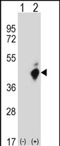
FAS Antibody (C-term Y291)
Affinity Purified Rabbit Polyclonal Antibody (Pab)
- 产品详情
- 实验流程
- 背景知识
Application
| WB, E |
|---|---|
| Primary Accession | P25445 |
| Other Accession | NP_690610.1, NP_690611.1, NP_000034.1 |
| Reactivity | Human |
| Host | Rabbit |
| Clonality | Polyclonal |
| Isotype | Rabbit IgG |
| Calculated MW | 37732 Da |
| Antigen Region | 269-298 aa |
| Gene ID | 355 |
|---|---|
| Other Names | Tumor necrosis factor receptor superfamily member 6, Apo-1 antigen, Apoptosis-mediating surface antigen FAS, FASLG receptor, CD95, FAS, APT1, FAS1, TNFRSF6 |
| Target/Specificity | This FAS antibody is generated from rabbits immunized with a KLH conjugated synthetic peptide between 269-298 amino acids from the C-terminal region of human FAS. |
| Dilution | WB~~1:1000 E~~Use at an assay dependent concentration. |
| Format | Purified polyclonal antibody supplied in PBS with 0.09% (W/V) sodium azide. This antibody is purified through a protein A column, followed by peptide affinity purification. |
| Storage | Maintain refrigerated at 2-8°C for up to 2 weeks. For long term storage store at -20°C in small aliquots to prevent freeze-thaw cycles. |
| Precautions | FAS Antibody (C-term Y291) is for research use only and not for use in diagnostic or therapeutic procedures. |
| Name | FAS |
|---|---|
| Synonyms | APT1, FAS1, TNFRSF6 |
| Function | Receptor for TNFSF6/FASLG. The adapter molecule FADD recruits caspase CASP8 to the activated receptor. The resulting death-inducing signaling complex (DISC) performs CASP8 proteolytic activation which initiates the subsequent cascade of caspases (aspartate-specific cysteine proteases) mediating apoptosis. FAS-mediated apoptosis may have a role in the induction of peripheral tolerance, in the antigen- stimulated suicide of mature T-cells, or both. The secreted isoforms 2 to 6 block apoptosis (in vitro). |
| Cellular Location | [Isoform 1]: Cell membrane; Single-pass type I membrane protein. Membrane raft [Isoform 3]: Secreted. [Isoform 5]: Secreted. |
| Tissue Location | Isoform 1 and isoform 6 are expressed at equal levels in resting peripheral blood mononuclear cells. After activation there is an increase in isoform 1 and decrease in the levels of isoform 6. |
For Research Use Only. Not For Use In Diagnostic Procedures.
Provided below are standard protocols that you may find useful for product applications.
BACKGROUND
The protein encoded by this gene is a member of the TNF-receptor superfamily. This receptor contains a death domain. It has been shown to play a central role in the physiological regulation of programmed cell death, and has been implicated in the pathogenesis of various malignancies and diseases of the immune system. The interaction of this receptor with its ligand allows the formation of a death-inducing signaling complex that includes Fas-associated death domain protein (FADD), caspase 8, and caspase 10. The autoproteolytic processing of the caspases in the complex triggers a downstream caspase cascade, and leads to apoptosis. This receptor has been also shown to activate NF-kappaB, MAPK3/ERK1, and MAPK8/JNK, and is found to be involved in transducing the proliferating signals in normal diploid fibroblast and T cells. At least eight alternatively spliced transcript variants have been described, some of which are candidates for nonsense-mediated decay (NMD). The isoforms lacking the transmembrane domain may negatively regulate the apoptosis mediated by the full length isoform.
REFERENCES
Cao, Y., et al. Mol. Carcinog. 49(11):944-950(2010)
Glavan, B.J., et al. Am. J. Respir. Crit. Care Med. (2010) In press :
Gizinger, O.A., et al. Vopr Kurortol Fizioter Lech Fiz Kult 3, 29-31 (2010) :
Dubikov, A.I., et al. Scand. J. Rheumatol. 39(5):368-372(2010)
Chakrabandhu, K., et al. EMBO J. 26(1):209-220(2007)
终于等到您。ABCEPTA(百远生物)抗体产品。
点击下方“我要评价 ”按钮提交您的反馈信息,您的反馈和评价是我们最宝贵的财富之一,
我们将在1-3个工作日内处理您的反馈信息。
如有疑问,联系:0512-88856768 tech-china@abcepta.com.


