


 癌症的基本特征包括细胞增殖、血管生成、迁移、凋亡逃避机制和细胞永生等。找到癌症发生过程中这些通路的关键标记物和对应的抗体用于检测至关重要。
 为您推荐一个泛素化位点预测神器——泛素化分析工具,可以为您的蛋白的泛素化位点作出预测和评分。
 细胞自噬受体图形绘图工具为你的蛋白的细胞受体结合位点作出预测和评分,识别结合到自噬通路中的蛋白是非常重要的,便于让我们理解自噬在正常生理、病理过程中的作用,如发育、细胞分化、神经退化性疾病、压力条件下、感染和癌症。
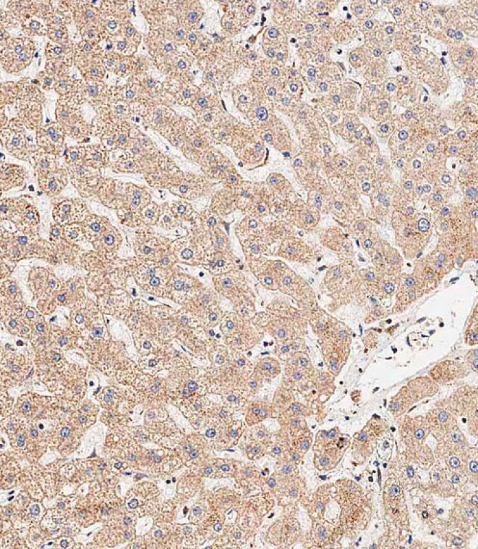
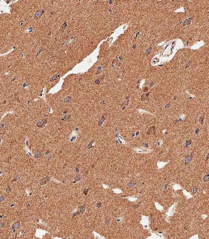
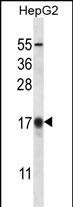
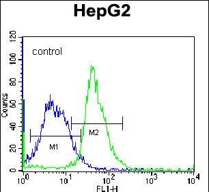
APG8b(MAP1LC3B) Antibody (N-term T29)
Affinity Purified Rabbit Polyclonal Antibody (Pab)
- 产品详情
- 实验流程
- 背景知识
Application
| IHC-P-Leica, WB, FC, E |
|---|---|
| Primary Accession | Q9GZQ8 |
| Other Accession | A6NCE7, Q62625, Q9CQV6, O41515, NP_073729.1 |
| Reactivity | Human, Rat, Mouse |
| Predicted | Rat, Bovine |
| Host | Rabbit |
| Clonality | Polyclonal |
| Isotype | Rabbit IgG |
| Calculated MW | 14688 Da |
| Antigen Region | 9-33 aa |
| Gene ID | 81631 |
|---|---|
| Other Names | Microtubule-associated proteins 1A/1B light chain 3B, Autophagy-related protein LC3 B, Autophagy-related ubiquitin-like modifier LC3 B, MAP1 light chain 3-like protein 2, MAP1A/MAP1B light chain 3 B, MAP1A/MAP1B LC3 B, Microtubule-associated protein 1 light chain 3 beta, MAP1LC3B, MAP1ALC3 |
| Target/Specificity | This APG8b(MAP1LC3B) antibody is generated from rabbits immunized with a KLH conjugated synthetic peptide between 9-33 amino acids from the N-terminal region of human APG8b(MAP1LC3B). |
| Dilution | IHC-P-Leica~~1:500 WB~~1:1000 FC~~1:10~50 E~~Use at an assay dependent concentration. |
| Format | Purified polyclonal antibody supplied in PBS with 0.09% (W/V) sodium azide. This antibody is purified through a protein A column, followed by peptide affinity purification. |
| Storage | Maintain refrigerated at 2-8°C for up to 2 weeks. For long term storage store at -20°C in small aliquots to prevent freeze-thaw cycles. |
| Precautions | APG8b(MAP1LC3B) Antibody (N-term T29) is for research use only and not for use in diagnostic or therapeutic procedures. |
| Name | MAP1LC3B (HGNC:13352) |
|---|---|
| Synonyms | MAP1ALC3 |
| Function | Ubiquitin-like modifier involved in formation of autophagosomal vacuoles (autophagosomes) (PubMed:20418806, PubMed:23209295, PubMed:28017329). Plays a role in mitophagy which contributes to regulate mitochondrial quantity and quality by eliminating the mitochondria to a basal level to fulfill cellular energy requirements and preventing excess ROS production (PubMed:23209295, PubMed:28017329). In response to cellular stress and upon mitochondria fission, binds C-18 ceramides and anchors autophagolysosomes to outer mitochondrial membranes to eliminate damaged mitochondria (PubMed:22922758). While LC3s are involved in elongation of the phagophore membrane, the GABARAP/GATE-16 subfamily is essential for a later stage in autophagosome maturation (PubMed:20418806, PubMed:23209295, PubMed:28017329). Promotes primary ciliogenesis by removing OFD1 from centriolar satellites via the autophagic pathway (PubMed:24089205). Through its interaction with the reticulophagy receptor TEX264, participates in the remodeling of subdomains of the endoplasmic reticulum into autophagosomes upon nutrient stress, which then fuse with lysosomes for endoplasmic reticulum turnover (PubMed:31006537, PubMed:31006538). Upon nutrient stress, directly recruits cofactor JMY to the phagophore membrane surfaces and promotes JMY's actin nucleation activity and autophagosome biogenesis during autophagy (PubMed:30420355). |
| Cellular Location | Cytoplasmic vesicle, autophagosome membrane; Lipid-anchor Endomembrane system; Lipid-anchor Mitochondrion membrane; Lipid-anchor. Cytoplasm, cytoskeleton {ECO:0000250|UniProtKB:Q9CQV6}. Cytoplasmic vesicle. Note=LC3-II binds to the autophagic membranes. LC3-II localizes with the mitochondrial inner membrane during Parkin-mediated mitophagy (PubMed:28017329). Also localizes to discrete punctae along the ciliary axoneme |
| Tissue Location | Most abundant in heart, brain, skeletal muscle and testis. Little expression observed in liver |
For Research Use Only. Not For Use In Diagnostic Procedures.
Provided below are standard protocols that you may find useful for product applications.
BACKGROUND
The product of this gene is a subunit of neuronal microtubule-associated MAP1A and MAP1B proteins, which are involved in microtubule assembly and important for neurogenesis. Studies on the rat homolog implicate a role for this gene in autophagy, a process that involves the bulk degradation of cytoplasmic component.
REFERENCES
Rouschop, K.M., et al. J. Clin. Invest. 120(1):127-141(2010)
Kirkin, V., et al. Mol. Cell 33(4):505-516(2009)
Othman, E.Q., et al. J. Clin. Lab. Anal. 23(4):249-258(2009)
Liu, Q., et al. Ai Zheng 27(1):25-29(2008)
Komatsu, M., et al. Cell 131(6):1149-1163(2007)
终于等到您。ABCEPTA(百远生物)抗体产品。
点击下方“我要评价 ”按钮提交您的反馈信息,您的反馈和评价是我们最宝贵的财富之一,
我们将在1-3个工作日内处理您的反馈信息。
如有疑问,联系:0512-88856768 tech-china@abcepta.com.


