


 癌症的基本特征包括细胞增殖、血管生成、迁移、凋亡逃避机制和细胞永生等。找到癌症发生过程中这些通路的关键标记物和对应的抗体用于检测至关重要。
 为您推荐一个泛素化位点预测神器——泛素化分析工具,可以为您的蛋白的泛素化位点作出预测和评分。
 细胞自噬受体图形绘图工具为你的蛋白的细胞受体结合位点作出预测和评分,识别结合到自噬通路中的蛋白是非常重要的,便于让我们理解自噬在正常生理、病理过程中的作用,如发育、细胞分化、神经退化性疾病、压力条件下、感染和癌症。
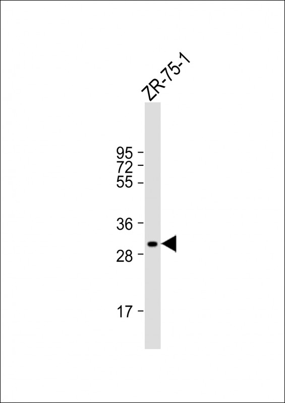
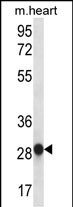
MYOGENIN Antibody (N-term)
Affinity Purified Rabbit Polyclonal Antibody (Pab)
- 产品详情
- 实验流程
- 背景知识
Application
| WB, E |
|---|---|
| Primary Accession | P15173 |
| Other Accession | NP_002470.2 |
| Reactivity | Human, Mouse |
| Host | Rabbit |
| Clonality | Polyclonal |
| Isotype | Rabbit IgG |
| Calculated MW | 25037 Da |
| Antigen Region | 1-30 aa |
| Gene ID | 4656 |
|---|---|
| Other Names | Myogenin, Class C basic helix-loop-helix protein 3, bHLHc3, Myogenic factor 4, Myf-4, MYOG, BHLHC3, MYF4 |
| Target/Specificity | This MYOGENIN antibody is generated from rabbits immunized with a KLH conjugated synthetic peptide between 1-30 amino acids from the N-terminal region of human MYOGENIN. |
| Dilution | WB~~1:1000 E~~Use at an assay dependent concentration. |
| Format | Purified polyclonal antibody supplied in PBS with 0.09% (W/V) sodium azide. This antibody is purified through a protein A column, followed by peptide affinity purification. |
| Storage | Maintain refrigerated at 2-8°C for up to 2 weeks. For long term storage store at -20°C in small aliquots to prevent freeze-thaw cycles. |
| Precautions | MYOGENIN Antibody (N-term) is for research use only and not for use in diagnostic or therapeutic procedures. |
| Name | MYOG |
|---|---|
| Synonyms | BHLHC3, MYF4 |
| Function | Acts as a transcriptional activator that promotes transcription of muscle-specific target genes and plays a role in muscle differentiation, cell cycle exit and muscle atrophy. Essential for the development of functional embryonic skeletal fiber muscle differentiation. However is dispensable for postnatal skeletal muscle growth; phosphorylation by CAMK2G inhibits its transcriptional activity in respons to muscle activity. Required for the recruitment of the FACT complex to muscle-specific promoter regions, thus promoting gene expression initiation. During terminal myoblast differentiation, plays a role as a strong activator of transcription at loci with an open chromatin structure previously initiated by MYOD1. Together with MYF5 and MYOD1, co-occupies muscle-specific gene promoter core regions during myogenesis. Also cooperates with myocyte-specific enhancer factor MEF2D and BRG1-dependent recruitment of SWI/SNF chromatin- remodeling enzymes to alter chromatin structure at myogenic late gene promoters. Facilitates cell cycle exit during terminal muscle differentiation through the up-regulation of miR-20a expression, which in turn represses genes involved in cell cycle progression. Binds to the E-box containing (E1) promoter region of the miR-20a gene. Also plays a role in preventing reversal of muscle cell differentiation. Contributes to the atrophy-related gene expression in adult denervated muscles. Induces fibroblasts to differentiate into myoblasts (By similarity). |
| Cellular Location | Nucleus. Note=Recruited to late myogenic gene promoter regulatory sequences with SMARCA4/BRG1/BAF190A and SWI/SNF chromatin-remodeling enzymes to promote chromatin-remodeling and transcription initiation in developing embryos. |
For Research Use Only. Not For Use In Diagnostic Procedures.
Provided below are standard protocols that you may find useful for product applications.
BACKGROUND
Myogenin is a muscle-specific transcription factor that can induce myogenesis in a variety of cell types in tissue culture. It is a member of a large family of proteins related by sequence homology, the helix-loop-helix (HLH) proteins. It is essential for the development of functional skeletal muscle. [provided by RefSeq].
REFERENCES
Gao, X., et al. J. Cell. Biochem. 110(1):162-170(2010)
Yerges, L.M., et al. J. Bone Miner. Res. 24(12):2039-2049(2009)
Ramamoorthy, S., et al. Am. J. Physiol. Endocrinol. Metab. 297 (2), E392-E401 (2009) :
Nanni, P., et al. Mol. Cancer Ther. 8(4):754-761(2009)
Gong, C., et al. Genes Dev. 23(1):54-66(2009)
终于等到您。ABCEPTA(百远生物)抗体产品。
点击下方“我要评价 ”按钮提交您的反馈信息,您的反馈和评价是我们最宝贵的财富之一,
我们将在1-3个工作日内处理您的反馈信息。
如有疑问,联系:0512-88856768 tech-china@abcepta.com.


