


 癌症的基本特征包括细胞增殖、血管生成、迁移、凋亡逃避机制和细胞永生等。找到癌症发生过程中这些通路的关键标记物和对应的抗体用于检测至关重要。
 为您推荐一个泛素化位点预测神器——泛素化分析工具,可以为您的蛋白的泛素化位点作出预测和评分。
 细胞自噬受体图形绘图工具为你的蛋白的细胞受体结合位点作出预测和评分,识别结合到自噬通路中的蛋白是非常重要的,便于让我们理解自噬在正常生理、病理过程中的作用,如发育、细胞分化、神经退化性疾病、压力条件下、感染和癌症。
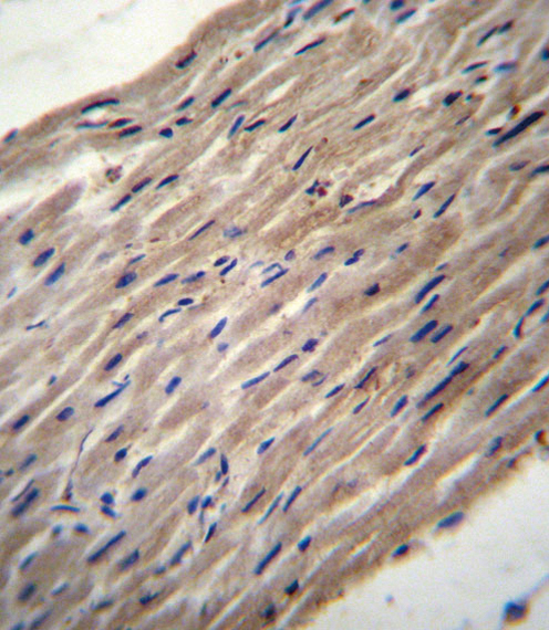
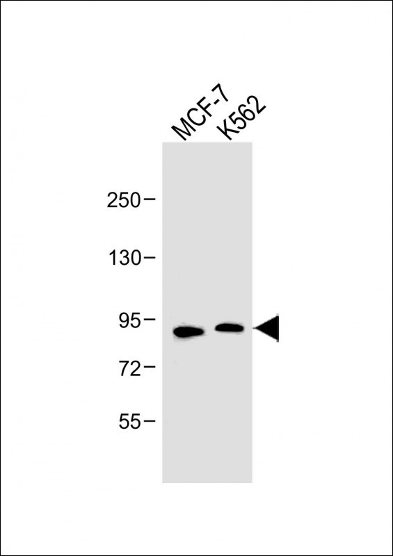
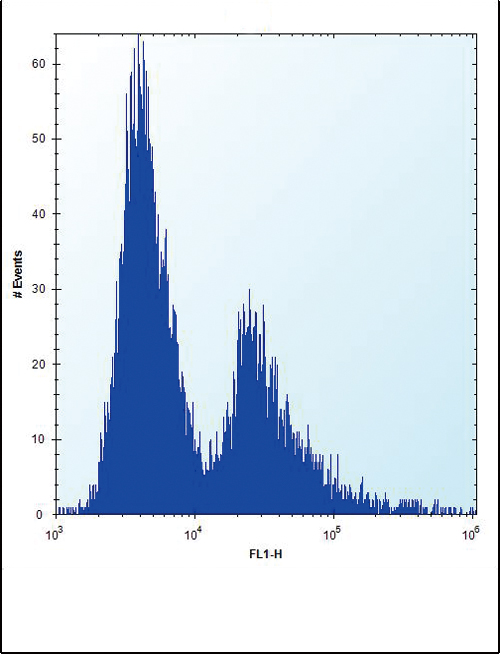
PLOD1 Antibody (N-term)
Affinity Purified Rabbit Polyclonal Antibody (Pab)
- 产品详情
- 文献引用 : 3
- 实验流程
- 背景知识
Application
| WB, IHC-P, FC, E |
|---|---|
| Primary Accession | Q02809 |
| Other Accession | Q9R0E2, NP_000293.2 |
| Reactivity | Human, Rat, Mouse |
| Predicted | Mouse |
| Host | Rabbit |
| Clonality | Polyclonal |
| Isotype | Rabbit IgG |
| Calculated MW | 83550 Da |
| Antigen Region | 66-94 aa |
| Gene ID | 5351 |
|---|---|
| Other Names | Procollagen-lysine, 2-oxoglutarate 5-dioxygenase 1, Lysyl hydroxylase 1, LH1, PLOD1, LLH, PLOD |
| Target/Specificity | This PLOD1 antibody is generated from rabbits immunized with a KLH conjugated synthetic peptide between 66-94 amino acids from the N-terminal region of human PLOD1. |
| Dilution | WB~~1:1000 IHC-P~~1:100~500 FC~~1:10~50 E~~Use at an assay dependent concentration. |
| Format | Purified polyclonal antibody supplied in PBS with 0.05% (V/V) Proclin 300. This antibody is purified through a protein A column, followed by peptide affinity purification. |
| Storage | Maintain refrigerated at 2-8°C for up to 2 weeks. For long term storage store at -20°C in small aliquots to prevent freeze-thaw cycles. |
| Precautions | PLOD1 Antibody (N-term) is for research use only and not for use in diagnostic or therapeutic procedures. |
| Name | PLOD1 |
|---|---|
| Synonyms | LLH, PLOD |
| Function | Part of a complex composed of PLOD1, P3H3 and P3H4 that catalyzes hydroxylation of lysine residues in collagen alpha chains and is required for normal assembly and cross-linkling of collagen fibrils (By similarity). Forms hydroxylysine residues in -Xaa-Lys- Gly- sequences in collagens (PubMed:10686424, PubMed:15854030, PubMed:8621606). These hydroxylysines serve as sites of attachment for carbohydrate units and are essential for the stability of the intermolecular collagen cross-links (Probable). |
| Cellular Location | Rough endoplasmic reticulum membrane; Peripheral membrane protein; Lumenal side |
For Research Use Only. Not For Use In Diagnostic Procedures.

Provided below are standard protocols that you may find useful for product applications.
BACKGROUND
Lysyl hydroxylase is a membrane-bound homodimeric protein localized to the cisternae of the endoplasmic reticulum. The enzyme (cofactors iron and ascorbate) catalyzes the hydroxylation of lysyl residues in collagen-like peptides. The resultant hydroxylysyl groups are attachment sites for carbohydrates in collagen and thus are critical for the stability of intermolecular crosslinks. Some patients with Ehlers-Danlos syndrome type VI have deficiencies in lysyl hydroxylase activity.
REFERENCES
Johnatty, S.E., et al. PLoS Genet. 6 (7), E1001016 (2010) :
Huang, Q.Y., et al. Bone 44(5):984-988(2009)
Yamada, Y., et al. Int. J. Mol. Med. 19(5):791-801(2007)
Tasker, P.N., et al. Osteoporos Int 17(7):1078-1085(2006)
Giunta, C., et al. Mol. Genet. Metab. 86 (1-2), 269-276 (2005) :
终于等到您。ABCEPTA(百远生物)抗体产品。
点击下方“我要评价 ”按钮提交您的反馈信息,您的反馈和评价是我们最宝贵的财富之一,
我们将在1-3个工作日内处理您的反馈信息。
如有疑问,联系:0512-88856768 tech-china@abcepta.com.


