


 癌症的基本特征包括细胞增殖、血管生成、迁移、凋亡逃避机制和细胞永生等。找到癌症发生过程中这些通路的关键标记物和对应的抗体用于检测至关重要。
 为您推荐一个泛素化位点预测神器——泛素化分析工具,可以为您的蛋白的泛素化位点作出预测和评分。
 细胞自噬受体图形绘图工具为你的蛋白的细胞受体结合位点作出预测和评分,识别结合到自噬通路中的蛋白是非常重要的,便于让我们理解自噬在正常生理、病理过程中的作用,如发育、细胞分化、神经退化性疾病、压力条件下、感染和癌症。
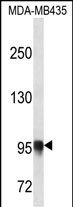
PLOD3 Antibody (N-term)
Affinity Purified Rabbit Polyclonal Antibody (Pab)
- 产品详情
- 实验流程
- 背景知识
Application
| WB, E |
|---|---|
| Primary Accession | O60568 |
| Other Accession | NP_001075.1 |
| Reactivity | Human |
| Host | Rabbit |
| Clonality | Polyclonal |
| Isotype | Rabbit IgG |
| Calculated MW | 84785 Da |
| Antigen Region | 78-105 aa |
| Gene ID | 8985 |
|---|---|
| Other Names | Procollagen-lysine, 2-oxoglutarate 5-dioxygenase 3, Lysyl hydroxylase 3, LH3, PLOD3 |
| Target/Specificity | This PLOD3 antibody is generated from rabbits immunized with a KLH conjugated synthetic peptide between 78-105 amino acids from the N-terminal region of human PLOD3. |
| Dilution | WB~~1:1000 E~~Use at an assay dependent concentration. |
| Format | Purified polyclonal antibody supplied in PBS with 0.09% (W/V) sodium azide. This antibody is purified through a protein A column, followed by peptide affinity purification. |
| Storage | Maintain refrigerated at 2-8°C for up to 2 weeks. For long term storage store at -20°C in small aliquots to prevent freeze-thaw cycles. |
| Precautions | PLOD3 Antibody (N-term) is for research use only and not for use in diagnostic or therapeutic procedures. |
| Name | PLOD3 |
|---|---|
| Function | Multifunctional enzyme that catalyzes a series of essential post-translational modifications on Lys residues in procollagen (PubMed:11956192, PubMed:12475640, PubMed:18298658, PubMed:18834968, PubMed:30089812). Plays a redundant role in catalyzing the formation of hydroxylysine residues in -Xaa-Lys-Gly- sequences in collagens (PubMed:11956192, PubMed:12475640, PubMed:18298658, PubMed:18834968, PubMed:30089812, PubMed:9582318, PubMed:9724729). Plays a redundant role in catalyzing the transfer of galactose onto hydroxylysine groups, giving rise to galactosyl 5-hydroxylysine (PubMed:12475640, PubMed:18298658, PubMed:18834968, PubMed:30089812). Has an essential role by catalyzing the subsequent transfer of glucose moieties, giving rise to 1,2-glucosylgalactosyl-5-hydroxylysine residues (PubMed:10934207, PubMed:11896059, PubMed:11956192, PubMed:12475640, PubMed:18298658, PubMed:18834968, PubMed:30089812). Catalyzes hydroxylation and glycosylation of Lys residues in the MBL1 collagen- like domain, giving rise to hydroxylysine and 1,2-glucosylgalactosyl-5- hydroxylysine residues (PubMed:25419660). Essential for normal biosynthesis and secretion of type IV collagens (Probable) (PubMed:18834968). Essential for normal formation of basement membranes (By similarity). |
| Cellular Location | Rough endoplasmic reticulum. Endoplasmic reticulum lumen. Endoplasmic reticulum membrane {ECO:0000250|UniProtKB:Q9R0E1}; Peripheral membrane protein {ECO:0000250|UniProtKB:Q9R0E1}; Lumenal side {ECO:0000250|UniProtKB:Q9R0E1}. Secreted Secreted, extracellular space {ECO:0000250|UniProtKB:Q9R0E1}. Note=The majority of the secreted protein is associated with the extracellular matrix. {ECO:0000250|UniProtKB:Q9R0E1} |
| Tissue Location | Ubiquitous (PubMed:9724729). Detected in heart, placenta and pancreas and at lower levels in lung, liver and skeletal muscle (PubMed:9582318, PubMed:9724729). |
For Research Use Only. Not For Use In Diagnostic Procedures.
Provided below are standard protocols that you may find useful for product applications.
BACKGROUND
The protein encoded by this gene is a membrane-bound homodimeric enzyme that is localized to the cisternae of the rough endoplasmic reticulum. The enzyme (cofactors iron and ascorbate) catalyzes the hydroxylation of lysyl residues in collagen-like peptides. The resultant hydroxylysyl groups are attachment sites for carbohydrates in collagen and thus are critical for the stability of intermolecular crosslinks. Some patients with Ehlers-Danlos syndrome type VIB have deficiencies in lysyl hydroxylase activity.
REFERENCES
Wang, C., et al. J. Cell. Mol. Med. 13(3):508-521(2009)
Salo, A.M., et al. Am. J. Hum. Genet. 83(4):495-503(2008)
Salo, A.M., et al. J. Cell. Physiol. 207(3):644-653(2006)
Wang, C., et al. Matrix Biol. 21(7):559-566(2002)
Rautavuoma, K., et al. J. Biol. Chem. 277(25):23084-23091(2002)
终于等到您。ABCEPTA(百远生物)抗体产品。
点击下方“我要评价 ”按钮提交您的反馈信息,您的反馈和评价是我们最宝贵的财富之一,
我们将在1-3个工作日内处理您的反馈信息。
如有疑问,联系:0512-88856768 tech-china@abcepta.com.


