


 癌症的基本特征包括细胞增殖、血管生成、迁移、凋亡逃避机制和细胞永生等。找到癌症发生过程中这些通路的关键标记物和对应的抗体用于检测至关重要。
 为您推荐一个泛素化位点预测神器——泛素化分析工具,可以为您的蛋白的泛素化位点作出预测和评分。
 细胞自噬受体图形绘图工具为你的蛋白的细胞受体结合位点作出预测和评分,识别结合到自噬通路中的蛋白是非常重要的,便于让我们理解自噬在正常生理、病理过程中的作用,如发育、细胞分化、神经退化性疾病、压力条件下、感染和癌症。
FKTN Antibody (Center)
Affinity Purified Rabbit Polyclonal Antibody (Pab)
- 产品详情
- 实验流程
- 背景知识
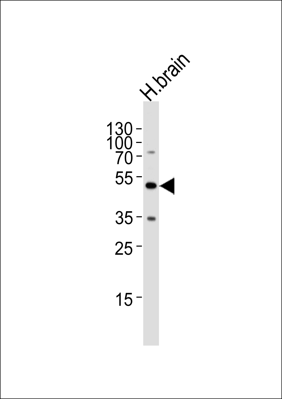
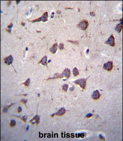
Application
| IHC-P, WB, E |
|---|---|
| Primary Accession | O75072 |
| Other Accession | Q60HG0, NP_001073270.1, NP_006722.2 |
| Reactivity | Human |
| Predicted | Monkey |
| Host | Rabbit |
| Clonality | Polyclonal |
| Isotype | Rabbit IgG |
| Calculated MW | 53724 Da |
| Antigen Region | 177-206 aa |
| Gene ID | 2218 |
|---|---|
| Other Names | Fukutin, 2---, Fukuyama-type congenital muscular dystrophy protein, FKTN, FCMD |
| Target/Specificity | This FKTN antibody is generated from rabbits immunized with a KLH conjugated synthetic peptide between 177-206 amino acids from the Central region of human FKTN. |
| Dilution | IHC-P~~1:100~500 WB~~1:1000 E~~Use at an assay dependent concentration. |
| Format | Purified polyclonal antibody supplied in PBS with 0.09% (W/V) sodium azide. This antibody is purified through a protein A column, followed by peptide affinity purification. |
| Storage | Maintain refrigerated at 2-8°C for up to 2 weeks. For long term storage store at -20°C in small aliquots to prevent freeze-thaw cycles. |
| Precautions | FKTN Antibody (Center) is for research use only and not for use in diagnostic or therapeutic procedures. |
| Name | FKTN (HGNC:3622) |
|---|---|
| Function | Catalyzes the transfer of a ribitol-phosphate from CDP- ribitol to the distal N-acetylgalactosamine of the phosphorylated O- mannosyl trisaccharide (N-acetylgalactosamine-beta-3-N- acetylglucosamine-beta-4-(phosphate-6-)mannose), a carbohydrate structure present in alpha-dystroglycan (DAG1) (PubMed:26923585, PubMed:27194101, PubMed:29477842). This constitutes the first step in the formation of the ribitol 5-phosphate tandem repeat which links the phosphorylated O-mannosyl trisaccharide to the ligand binding moiety composed of repeats of 3-xylosyl-alpha-1,3-glucuronic acid-beta-1 (PubMed:17034757, PubMed:25279699, PubMed:26923585, PubMed:27194101, PubMed:29477842). Required for normal location of POMGNT1 in Golgi membranes, and for normal POMGNT1 activity (PubMed:17034757). May interact with and reinforce a large complex encompassing the outside and inside of muscle membranes (PubMed:25279699). Could be involved in brain development (Probable). |
| Cellular Location | Golgi apparatus membrane; Single-pass type II membrane protein. Cytoplasm {ECO:0000250|UniProtKB:Q8R507}. Nucleus {ECO:0000250|UniProtKB:Q8R507}. Note=In retinal tissue, does not localize with the Golgi apparatus. {ECO:0000250|UniProtKB:Q8R507} |
| Tissue Location | Expressed in the retina (at protein level) (PubMed:29416295). Widely expressed with highest expression in brain, heart, pancreas and skeletal muscle (PubMed:11115853). Expressed at similar levels in control fetal and adult brain (PubMed:11115853) Expressed in migrating neurons, including Cajar-Retzius cells and adult cortical neurons, as well as hippocampal pyramidal cells and cerebellar Purkinje cells (PubMed:11115853). No expression observed in the glia limitans, the subpial astrocytes (which contribute to basement membrane formation) or other glial cells (PubMed:11115853) |
For Research Use Only. Not For Use In Diagnostic Procedures.
Provided below are standard protocols that you may find useful for product applications.
BACKGROUND
The protein encoded by this gene is a putative transmembrane protein that is localized to the cis-Golgi compartment, where it may be involved in the glycosylation of alpha-dystroglycan in skeletal muscle. The encoded protein is thought to be a glycosyltransferase and could play a role in brain development. Defects in this gene are a cause of Fukuyama-type congenital muscular dystrophy (FCMD), Walker-Warburg syndrome (WWS), limb-girdle muscular dystrophy type 2M (LGMD2M), and dilated cardiomyopathy type 1X (CMD1X). Alternatively spliced transcript variants have been found for this gene.
REFERENCES
Lim, B.C., et al. Neuromuscul. Disord. 20(8):524-530(2010)
Saredi, S., et al. Muscle Nerve 39(6):845-848(2009)
Chang, W., et al. Prenat. Diagn. 29(6):560-569(2009)
Mercuri, E., et al. Neurology 72(21):1802-1809(2009)
Puckett, R.L., et al. Neuromuscul. Disord. 19(5):352-356(2009)
终于等到您。ABCEPTA(百远生物)抗体产品。
点击下方“我要评价 ”按钮提交您的反馈信息,您的反馈和评价是我们最宝贵的财富之一,
我们将在1-3个工作日内处理您的反馈信息。
如有疑问,联系:0512-88856768 tech-china@abcepta.com.


