


 癌症的基本特征包括细胞增殖、血管生成、迁移、凋亡逃避机制和细胞永生等。找到癌症发生过程中这些通路的关键标记物和对应的抗体用于检测至关重要。
 为您推荐一个泛素化位点预测神器——泛素化分析工具,可以为您的蛋白的泛素化位点作出预测和评分。
 细胞自噬受体图形绘图工具为你的蛋白的细胞受体结合位点作出预测和评分,识别结合到自噬通路中的蛋白是非常重要的,便于让我们理解自噬在正常生理、病理过程中的作用,如发育、细胞分化、神经退化性疾病、压力条件下、感染和癌症。
CLN3 Antibody (Center)
Affinity Purified Rabbit Polyclonal Antibody (Pab)
- 产品详情
- 实验流程
- 背景知识
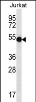
Application
| WB, E |
|---|---|
| Primary Accession | Q13286 |
| Other Accession | Q60HH0, NP_001035897.1, NP_000077.1 |
| Reactivity | Human |
| Predicted | Monkey |
| Host | Rabbit |
| Clonality | Polyclonal |
| Isotype | Rabbit IgG |
| Calculated MW | 47623 Da |
| Antigen Region | 235-263 aa |
| Gene ID | 1201 |
|---|---|
| Other Names | Battenin, Batten disease protein, Protein CLN3, CLN3, BTS |
| Target/Specificity | This CLN3 antibody is generated from rabbits immunized with a KLH conjugated synthetic peptide between 235-263 amino acids from the Central region of human CLN3. |
| Dilution | WB~~1:1000 E~~Use at an assay dependent concentration. |
| Format | Purified polyclonal antibody supplied in PBS with 0.09% (W/V) sodium azide. This antibody is purified through a protein A column, followed by peptide affinity purification. |
| Storage | Maintain refrigerated at 2-8°C for up to 2 weeks. For long term storage store at -20°C in small aliquots to prevent freeze-thaw cycles. |
| Precautions | CLN3 Antibody (Center) is for research use only and not for use in diagnostic or therapeutic procedures. |
| Name | CLN3 (HGNC:2074) |
|---|---|
| Synonyms | BTS |
| Function | Mediates microtubule-dependent, anterograde transport connecting the Golgi network, endosomes, autophagosomes, lysosomes and plasma membrane, and participates in several cellular processes such as regulation of lysosomal pH, lysosome protein degradation, receptor- mediated endocytosis, autophagy, transport of proteins and lipids from the TGN, apoptosis and synaptic transmission (PubMed:10924275, PubMed:15471887, PubMed:18317235, PubMed:18817525, PubMed:20850431, PubMed:22261744). Facilitates the proteins transport from trans-Golgi network (TGN)-to other membrane compartments such as transport of microdomain-associated proteins to the plasma membrane, IGF2R transport to the lysosome where it regulates the CTSD release leading to regulation of CTSD maturation and thereby APP intracellular processing (PubMed:10924275, PubMed:18817525). Moreover regulates CTSD activity in response to osmotic stress (PubMed:23840424, PubMed:28390177). Also binds galactosylceramide and transports it from the trans Golgi to the rafts, which may have immediate and downstream effects on cell survival by modulating ceramide synthesis (PubMed:18317235). At the plasma membrane, regulates actin-dependent events including filopodia formation, cell migration, and pinocytosis through ARF1-CDC42 pathway and also the cytoskeleton organization through interaction with MYH10 and fodrin leading to the regulation of the plasma membrane association of Na+, K+ ATPase complex (PubMed:20850431). Regulates synaptic transmission in the amygdala, hippocampus, and cerebellum through regulation of synaptic vesicles density and their proximity to active zones leading to modulation of short-term plasticity and age-dependent anxious behavior, learning and memory (By similarity). Regulates autophagic vacuoles (AVs) maturation by modulating the trafficking between endocytic and autophagolysosomal/lysosomal compartments, which involves vesicle fusion leading to regulation of degradation process (By similarity). Also participates in cellular homeostasis of compounds such as, water, ions, amino acids, proteins and lipids in several tissue namely in brain and kidney through regulation of their transport and synthesis (PubMed:17482562). |
| Cellular Location | Lysosome membrane; Multi-pass membrane protein. Late endosome. Lysosome. Golgi apparatus. Golgi apparatus membrane. Golgi apparatus, Golgi stack. Golgi apparatus, trans-Golgi network. Cell membrane Recycling endosome. Membrane raft. Membrane, caveola. Early endosome membrane. Synapse, synaptosome {ECO:0000250|UniProtKB:Q61124}. Late endosome membrane {ECO:0000250|UniProtKB:Q61124}. Cytoplasmic vesicle, autophagosome {ECO:0000250|UniProtKB:Q61124}. Note=CLN3 is not present in late endosomes/lysosomes in fibroblasts and neurons (PubMed:15240864) Trafficks from cell membrane to Golgi via endosomes (PubMed:15240864) Osmotic stress changes the subcellular localization of CLN3 (PubMed:23840424). Trafficks to intracellular compartments via the plasma membranet through AP3M1-dependent mechanisms (PubMed:14644441) Excluded from the synaptic vesicles (By similarity) {ECO:0000250|UniProtKB:Q61124, ECO:0000269|PubMed:14644441, ECO:0000269|PubMed:15240864, ECO:0000269|PubMed:23840424} |
| Tissue Location | Expressed in the cortical brain, pancreas, spleen, and testis with weaker expression in the peripheral nerve (at protein level). Highly expressed in gray matter (at protein level) |
For Research Use Only. Not For Use In Diagnostic Procedures.
Provided below are standard protocols that you may find useful for product applications.
BACKGROUND
This gene encodes a protein that is involved in lysosomal function. Mutations in this, as well as other neuronal ceroid-lipofuscinosis (CLN) genes, cause neurodegenerative diseases commonly known as Batten disease or collectively known as neuronal ceroid lipofuscinoses (NCLs). Many alternatively spliced transcript variants have been found for this gene.
REFERENCES
Adams, H.R., et al. Dev Med Child Neurol 52(7):637-643(2010)
Imielinski, M., et al. Nat. Genet. 41(12):1335-1340(2009)
Sarpong, A., et al. Clin. Genet. 76(1):38-45(2009)
Codlin, S., et al. J. Cell. Sci. 122 (PT 8), 1163-1173 (2009) :
Tuxworth, R.I., et al. Hum. Mol. Genet. 18(4):667-678(2009)
终于等到您。ABCEPTA(百远生物)抗体产品。
点击下方“我要评价 ”按钮提交您的反馈信息,您的反馈和评价是我们最宝贵的财富之一,
我们将在1-3个工作日内处理您的反馈信息。
如有疑问,联系:0512-88856768 tech-china@abcepta.com.


