


 癌症的基本特征包括细胞增殖、血管生成、迁移、凋亡逃避机制和细胞永生等。找到癌症发生过程中这些通路的关键标记物和对应的抗体用于检测至关重要。
 为您推荐一个泛素化位点预测神器——泛素化分析工具,可以为您的蛋白的泛素化位点作出预测和评分。
 细胞自噬受体图形绘图工具为你的蛋白的细胞受体结合位点作出预测和评分,识别结合到自噬通路中的蛋白是非常重要的,便于让我们理解自噬在正常生理、病理过程中的作用,如发育、细胞分化、神经退化性疾病、压力条件下、感染和癌症。
HtrA1 Antibody (C-term)
Purified Rabbit Polyclonal Antibody (Pab)

- 产品详情
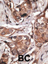
- 文献引用 : 1
- 实验流程
- 背景知识
Application
| WB, IHC-P, E |
|---|---|
| Primary Accession | Q92743 |
| Other Accession | Q9QZK5 |
| Reactivity | Human |
| Predicted | Rat |
| Host | Rabbit |
| Clonality | Polyclonal |
| Isotype | Rabbit IgG |
| Calculated MW | 51287 Da |
| Antigen Region | 381-412 aa |
| Gene ID | 5654 |
|---|---|
| Other Names | Serine protease HTRA1, 3421-, High-temperature requirement A serine peptidase 1, L56, Serine protease 11, HTRA1, HTRA, PRSS11 |
| Target/Specificity | This HtrA1 antibody is generated from rabbits immunized with a KLH conjugated synthetic peptide between 381-412 amino acids from the C-terminal region of human HtrA1. |
| Dilution | WB~~1:1000 IHC-P~~1:100~500 E~~Use at an assay dependent concentration. |
| Format | Purified polyclonal antibody supplied in PBS with 0.05% (V/V) Proclin 300. This antibody is prepared by Saturated Ammonium Sulfate (SAS) precipitation followed by dialysis against PBS. |
| Storage | Maintain refrigerated at 2-8°C for up to 2 weeks. For long term storage store at -20°C in small aliquots to prevent freeze-thaw cycles. |
| Precautions | HtrA1 Antibody (C-term) is for research use only and not for use in diagnostic or therapeutic procedures. |
| Name | HTRA1 |
|---|---|
| Synonyms | HTRA, PRSS11 |
| Function | Serine protease with a variety of targets, including extracellular matrix proteins such as fibronectin. HTRA1-generated fibronectin fragments further induce synovial cells to up-regulate MMP1 and MMP3 production. May also degrade proteoglycans, such as aggrecan, decorin and fibromodulin. Through cleavage of proteoglycans, may release soluble FGF-glycosaminoglycan complexes that promote the range and intensity of FGF signals in the extracellular space. Regulates the availability of insulin-like growth factors (IGFs) by cleaving IGF- binding proteins. Inhibits signaling mediated by TGF-beta family members. This activity requires the integrity of the catalytic site, although it is unclear whether TGF-beta proteins are themselves degraded. By acting on TGF-beta signaling, may regulate many physiological processes, including retinal angiogenesis and neuronal survival and maturation during development. Intracellularly, degrades TSC2, leading to the activation of TSC2 downstream targets. |
| Cellular Location | Cell membrane. Secreted Cytoplasm, cytosol. Note=Predominantly secreted (PubMed:15208355). Also found associated with the plasma membrane (PubMed:21297635). |
| Tissue Location | Widely expressed, with strongest expression in placenta (at protein level). Secreted by synovial fibroblasts. Up- regulated in osteoarthritis and rheumatoid arthritis synovial fluids and cartilage as compared with non-arthritic (at protein level) |
For Research Use Only. Not For Use In Diagnostic Procedures.

Provided below are standard protocols that you may find useful for product applications.
BACKGROUND
HtrA1 is a member of the trypsin family of serine proteases. This protein is a secreted enzyme that is proposed to regulate the availability of insulin-like growth factors (IGFs) by cleaving IGF-binding proteins. It has also been suggested to be a regulator of cell growth.
REFERENCES
Howes, N., et al., Clin Gastroenterol Hepatol 2(3):252-261 (2004).
Chien, J., et al., Oncogene 23(8):1636-1644 (2004).
Hu, S.I., et al., J. Biol. Chem. 273(51):34406-34412 (1998).
Zumbrunn, J., et al., Genomics 45(2):461-462 (1997).
Zumbrunn, J., et al., FEBS Lett. 398 (2-3), 187-192 (1996).
终于等到您。ABCEPTA(百远生物)抗体产品。
点击下方“我要评价 ”按钮提交您的反馈信息,您的反馈和评价是我们最宝贵的财富之一,
我们将在1-3个工作日内处理您的反馈信息。
如有疑问,联系:0512-88856768 tech-china@abcepta.com.


